
Photomicrography of Hydra - a model for studying regeneration and aging
by Dr. Robert Berdan
August 3, 2018

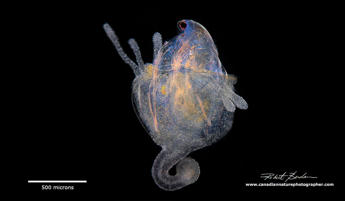
Hydra viridissima - the Green hydra with a bud. The green colour is due to endosymbiotic algae (Chlorella) living inside the hydra.

Diagram of Hydra -it is essentially a hollow tube with tentacles, has two layers of cells an outer ectoderm and an inner mesoderm (gastrodermis) of cells separated by a non-cellular mesoglea.

Above is a thin section through the hydra examined with an electron microscope. On the left is the thin layer of ectoderm separated by mesoglea and on the right are the columnar cells comprising the endoderm that face the gut. Most of the animal is composed of a hydroskeleton - open spaces filled with water. I did some research on Hydra when I was a graduate student and took this picture at the University of Western Ontario. In this article I revisit my old friend to take some pictures with my Axioscope microscope with DIC (differential interference contrast) microscopy as well as by bright field and darkfield microscopy. In this article I go a little deeper in depth then usual for those that might be interested in the biology of Hydra, otherwise I hope you will enjoy the pictures.
Introduction to Hydra
The name Hydra was derived from a serpentine water monster in Greek and Roman mythology, see painting of Hercules and the serpent below. The Hydra had poisonous breath and blood so virulent that even its scent was deadly. Later versions of the Hydra story add a regeneration feature to the monster: for every head chopped off, the Hydra would regrow two heads (read more at Wikipedia).

Hercules and the Lernaean Hydra painting - work is in the public domain. Source Wikipedia - Gustave Moreau 19th-century depiction of Hydra.
Research interests in Hydra living in pond water surround two interesting facts about this organism, its amazing ability to regenerate and unlike most other organisms it does not appear to age (Martínez 1998).
I revisited Hydra again recently when collecting pond water and found two species in my jars; one a smaller green hydra (Hydra viridissima) and a second larger brownish-translucent species. The Hydra attached to the bottom and sides of my plastic petri dish and hang down from the surface of the water film. They were also attached to many of the water plants (spiked water milfoil) I collected from a fish pond in the foothills of the rocky mountains near my home in Calgary.
Hydra are found in fresh water ponds usually attached to water-plants. They can sometimes be abundant and other times rare.
Identification of the different species of hydra is determined by three main criteria (Campbell 1983).
1. Presence or absence of symbiotic algae - H.viridissima or H. hadleyi
2. Arrangement of tentacles on buds - they can arise simultaneously, some arise early, tentacles can also be staggered in appearance.
3. Shape of isorhiza nematocyst - length to width ratio of Holotrichous isorhiza nematocyst (see below). There are 4 types of nematocysts. In my brown specimens the width was less than 1\2 the length so its likely to belong to the Oligactis group or Vulgaris group of Hydra but without observing bud tentacles I can't be sure which it is.

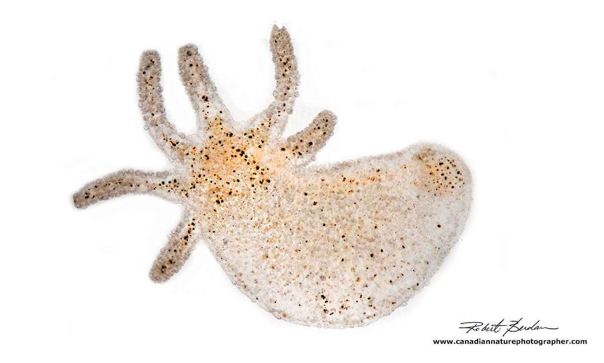
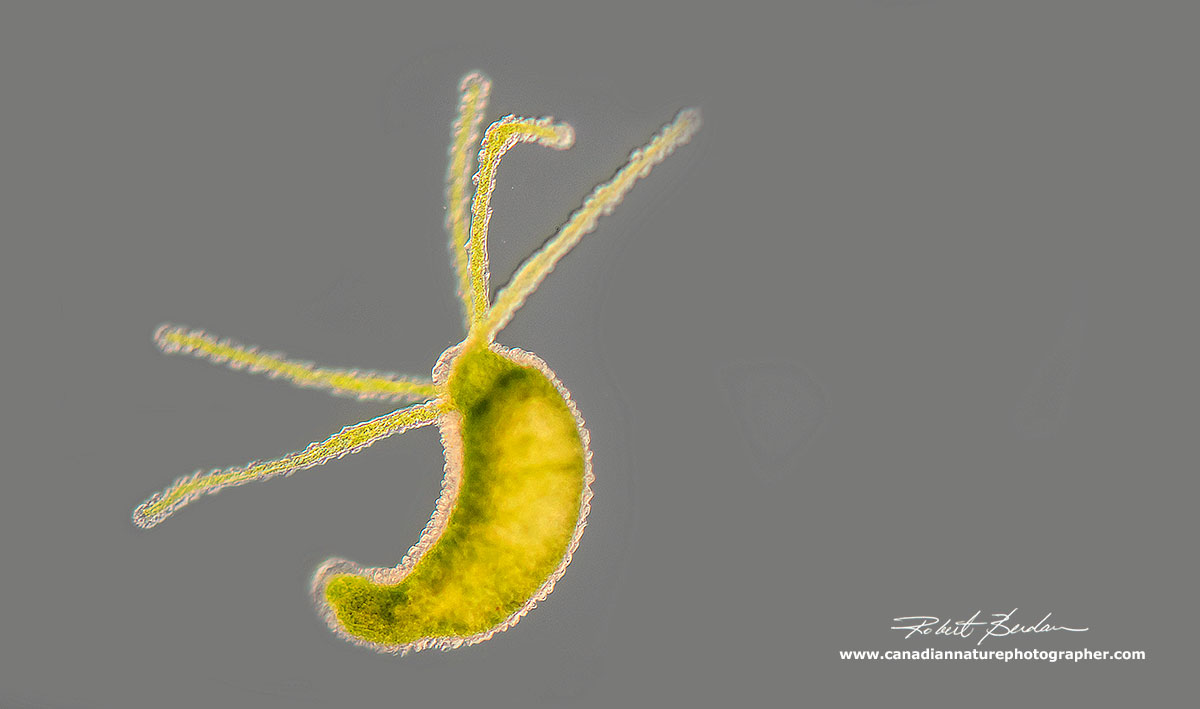
Hydra can range in size from 0.5 mm to 10 mm in length and their bodies are highly contractile as shown in the picture above of the same animal. Hydra viridissima.

Hydra by Darkfield microscopy at low magnification showing the body column and hydranth.
Hydra hydranth via Dark field microscopy shows many intracellular particles and granules.
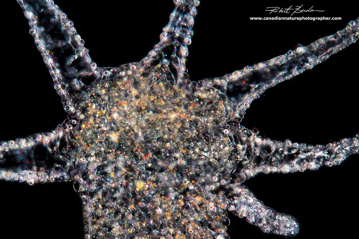
Low magnification picture of brown Hydra using bright field microscopy.

Hydra viridissima by bright field microscopy showing the numerous green algae inside.
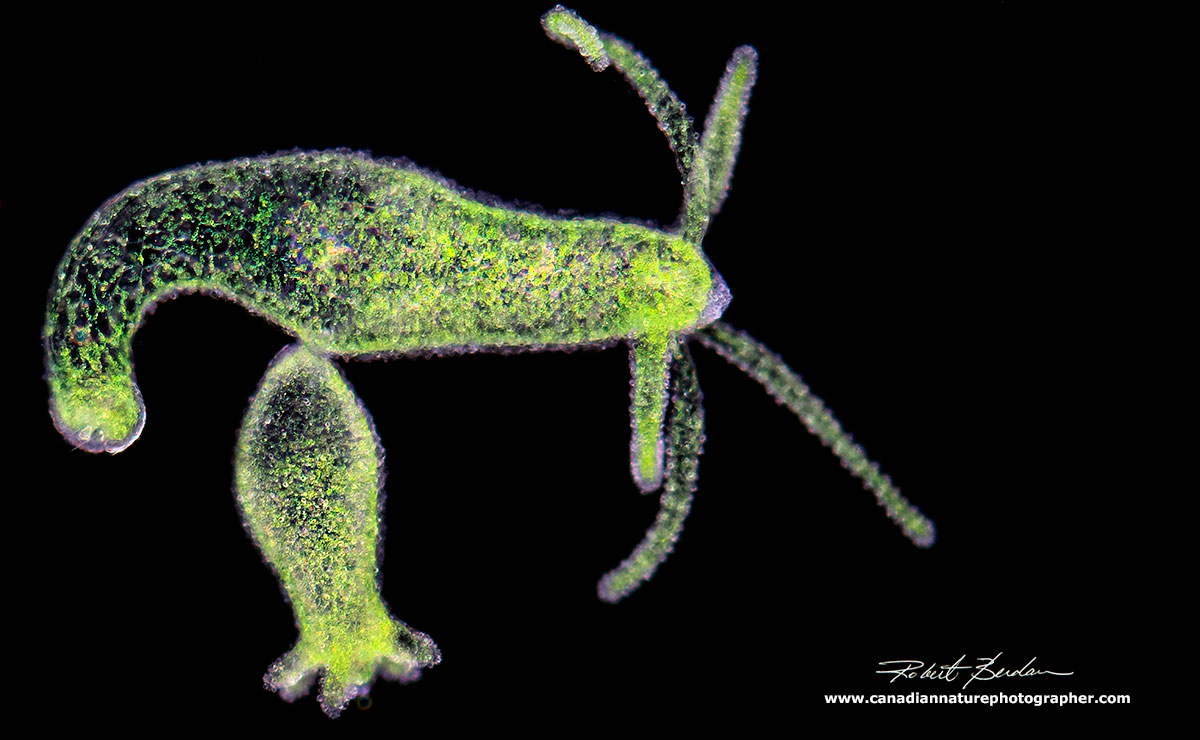
Hydra viridissima with bud note the green colour due to endosymbiotic algae (Chlorella).

Hydra viridissima contracted state. This hydra tends to be much smaller then my brown hydra.

Close up of the hydranth of Hydra viridissima with tentacles retracted - DIC microscopy.
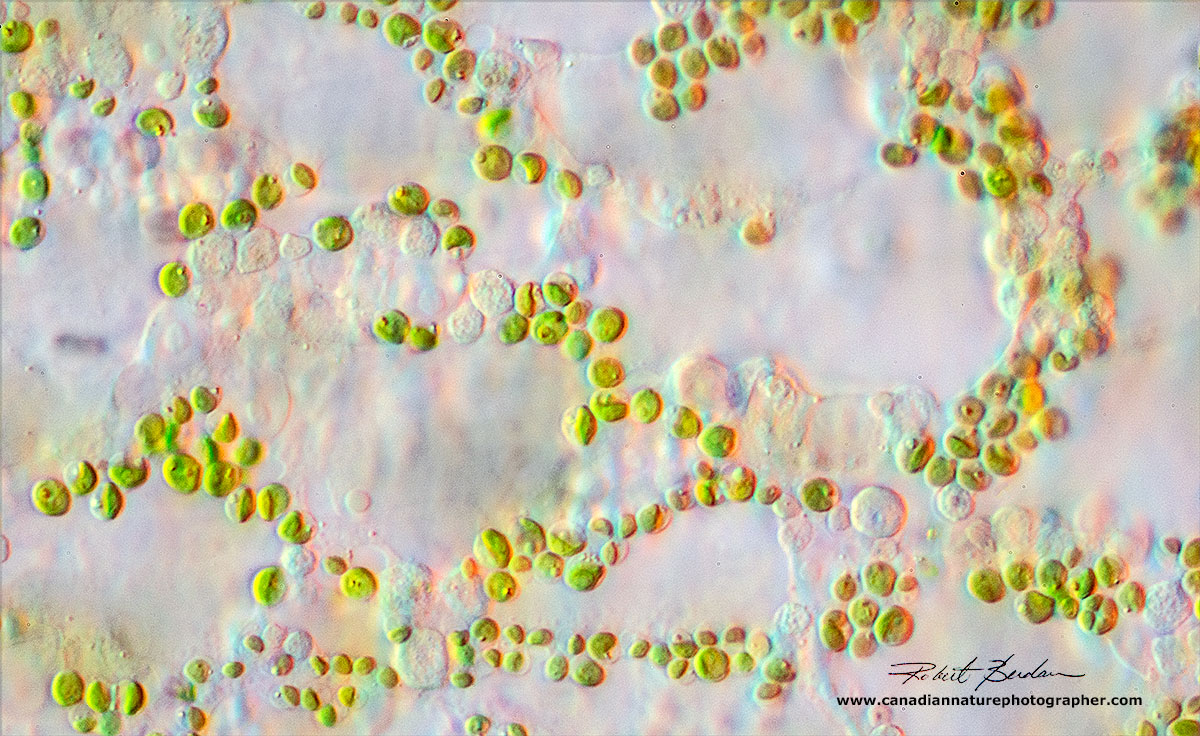
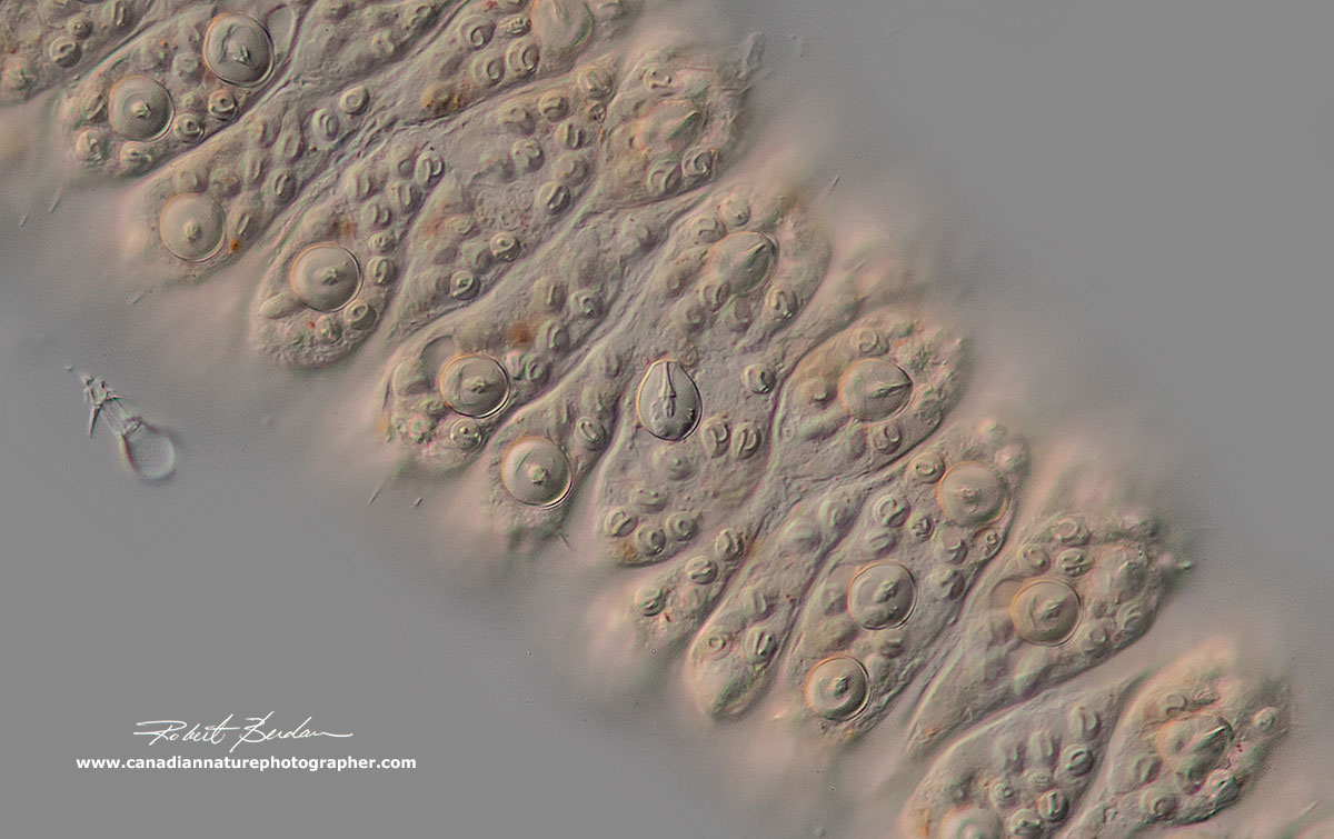
Close up of the body wall of Hydra showing the Chlorella cells inside, DIC microscopy.
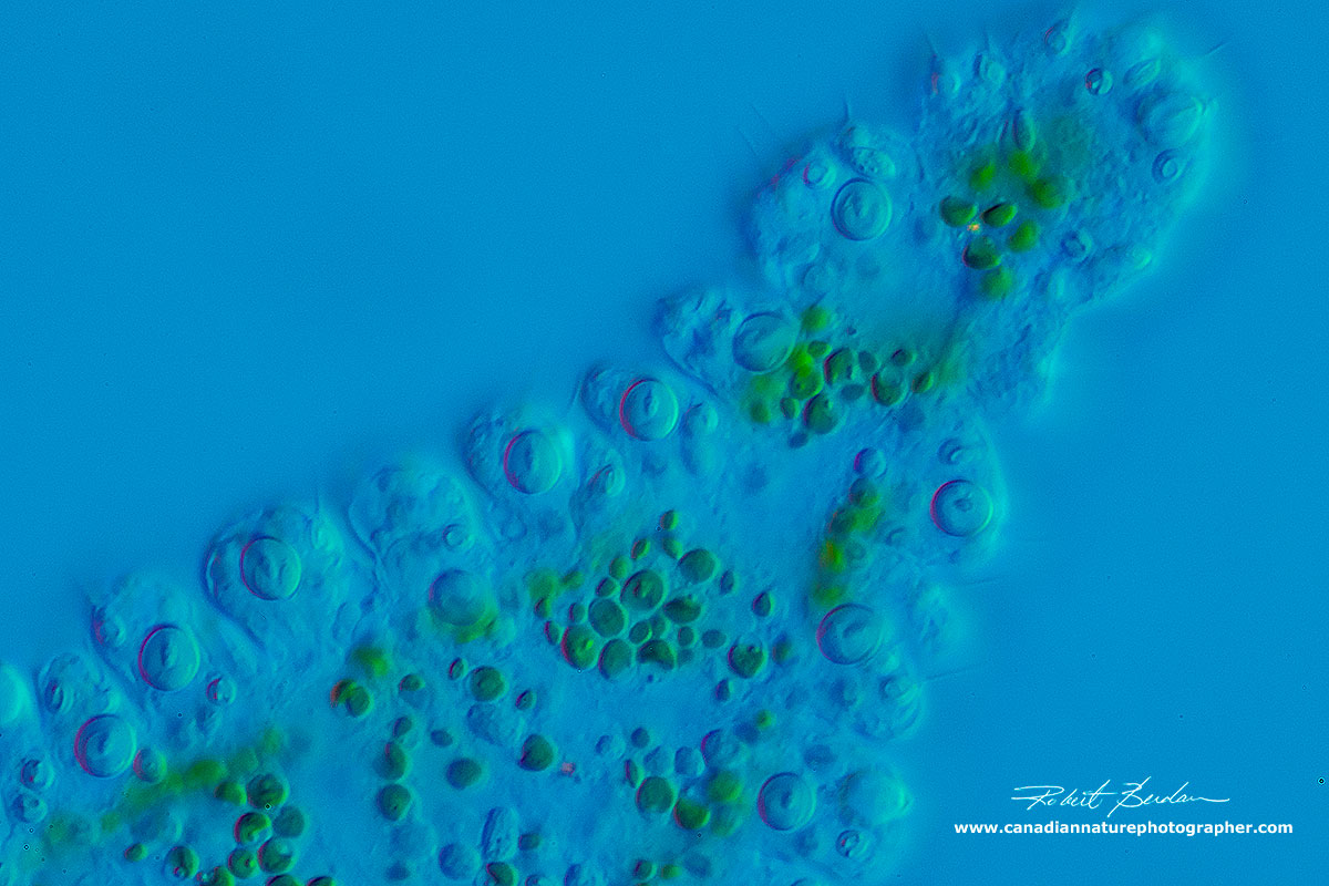
Hydra tentacle with Chlorella, on the outer edges are numerous nematocysts also called cnidoblasts (stinging cells). DIC microscopy.

Hydra hydranth - the mouth opens at the apical end. DIC microscopy

Hydra hydranth showing the tentacles which are packed with cnidoblasts - stinging cells. The clumps are sometimes referred to as cnidocyte batteries and were first described by Antony van Leeuweenhoek in 1702. Later Hydra was first studied in detail by Abraham Trembley (1720-1784) who noted Hydra's incredible regenerative capacity. Hydra can even regenerate from single cells which form re-aggregates after the tissues are dissociated. Hydra also shares 6071 genes with humans (humans have about 20,000 coding genes).
Hydra viridissima low magnification DIC microscopy. A single Hydra contains between 50,000 and 100, 000 cells and 3 distinct stem cell populations: 1) interstitial 2) ectodermal epithelial and 3) endodermal epithelial cells that give rise to about a dozen different cell types. These stem cells constantly self renew. The only other cells I am aware that do this are cancer cells. Among 259 human aging genes 207 (80%) were conserved in Hydra oligactis (Tomczyk et al 2014).

Hydra viridissima with a bud - DIC microscopy
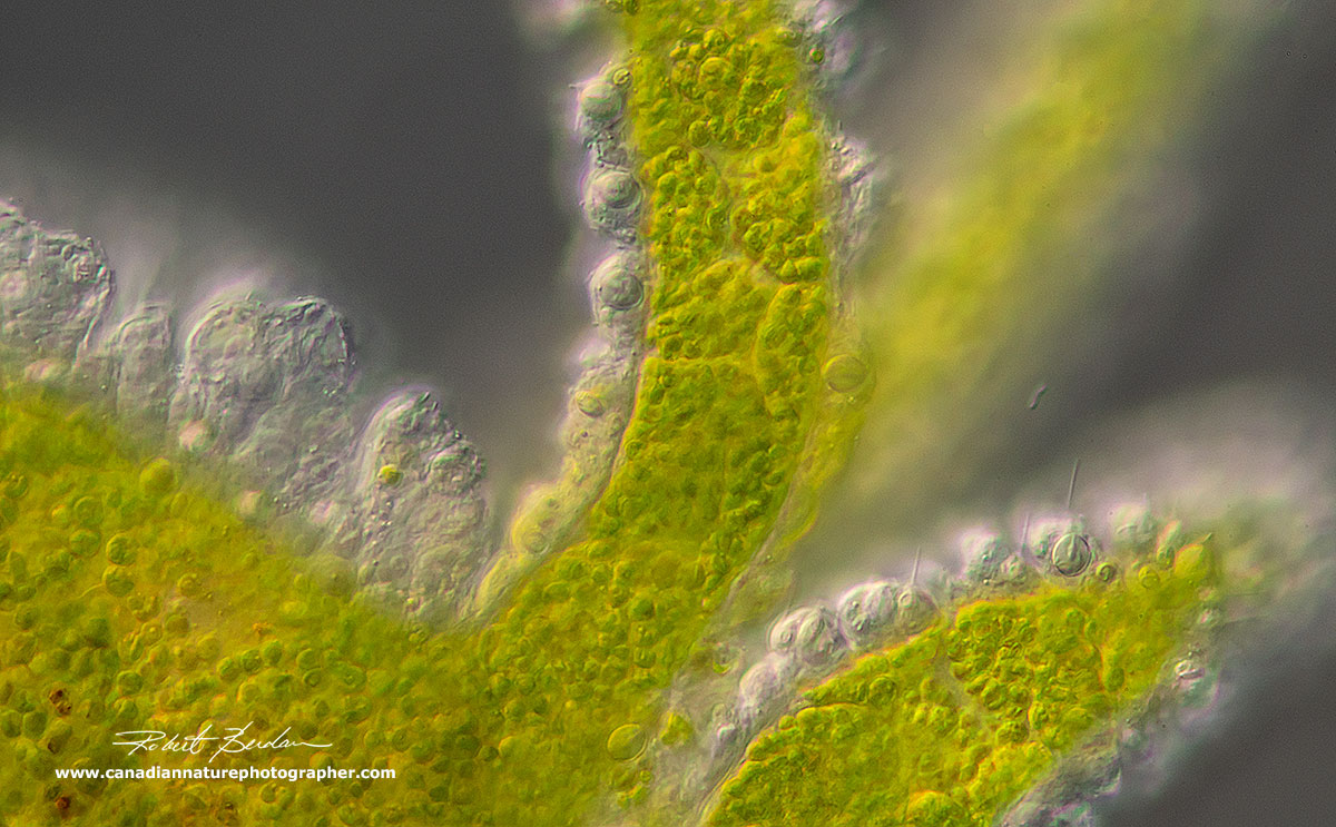
Hydra viridissima showing the tentacles and some of the cnidoblasts can be seen
Hyrda showing its great flexibility - DIC microscopy about 200X

Closeup of the Basal Disk of Hydra which it uses to attach to the substratum - DIC microscopy.
Cnidoblasts
A cnidocyte (also known as a cnidoblast or nematocyte) is an explosive cell containing one giant secretory organelle or cnida (plural cnidae) that defines the phylum Cnidaria (corals, sea anemones, hydrae, jellyfish, etc.). The cindocytes fire a dart-like thread containing a neurotoxin that paralyze prey. On freshwater hydra you are not likely to feel them as they are too small. These stinging cells are concentrated on the tentacles and look like miniature light bulbs with a thick coiled thread inside. Upon contact with prey the contents of the nematocyst are explosively discharged. they can also be triggered to fire with weak acids (e.g. vinegar). There are 4 types of cnidocyte found on Hydra (see below) and by measuring their length\width ratio these measurements can be used to identify some Hydra species.

Firing mechanism of hydra nematocyst Ref:Ruppert, E.E., Fox, R.S., and Barnes, R.D. (2004) Invertebrate Zoology (7th ed.), Brooks / Cole, pp. 111-124 - Philcha Wikipedia

Four types of nematocysts - one measures the length (L) and width (W) to identify the species.
(Modified from Campbell, 1983 & Deserti and Zamponi 2012)

Above is a picture of the surface of Hydra showing the 4 different types of nematocyst. To correctly identify the species one would measure the length and width to determine the ratios - refer to Campbell 1983 or Deserti and Zamponi 2012. In science these measurements are referred to as a morphometric analysis. Atrichuos isorhiza nematocysts are without spines and attach to the substratum in a manner like Batman's Batropes and can be used to pull them along. Desmoneme cnidoblast is used for prey attachment. Stenotele's, and the isorhiza cnidoblasts are able to pierce cuticle (Balasubramanian et al. 2012 Proteome of Hydra Nematocyst J. Biol. chem 287: 9672 PDF)

Hydra tentacle showing a discharged nematocyst. The circular and oval objects on the tentacles are nematocysts.

Electron micrograph of a developing cnidoblast - R. Berdan unpublished electron micrograph.
Cnidoblasts cover the Hydra's tentacles. DIC microscopy.

Cnidoblasts cover the Hydra's tentacles. DIC microscopy.

Cnidoblasts stained with Neutral red DIC microscopy.
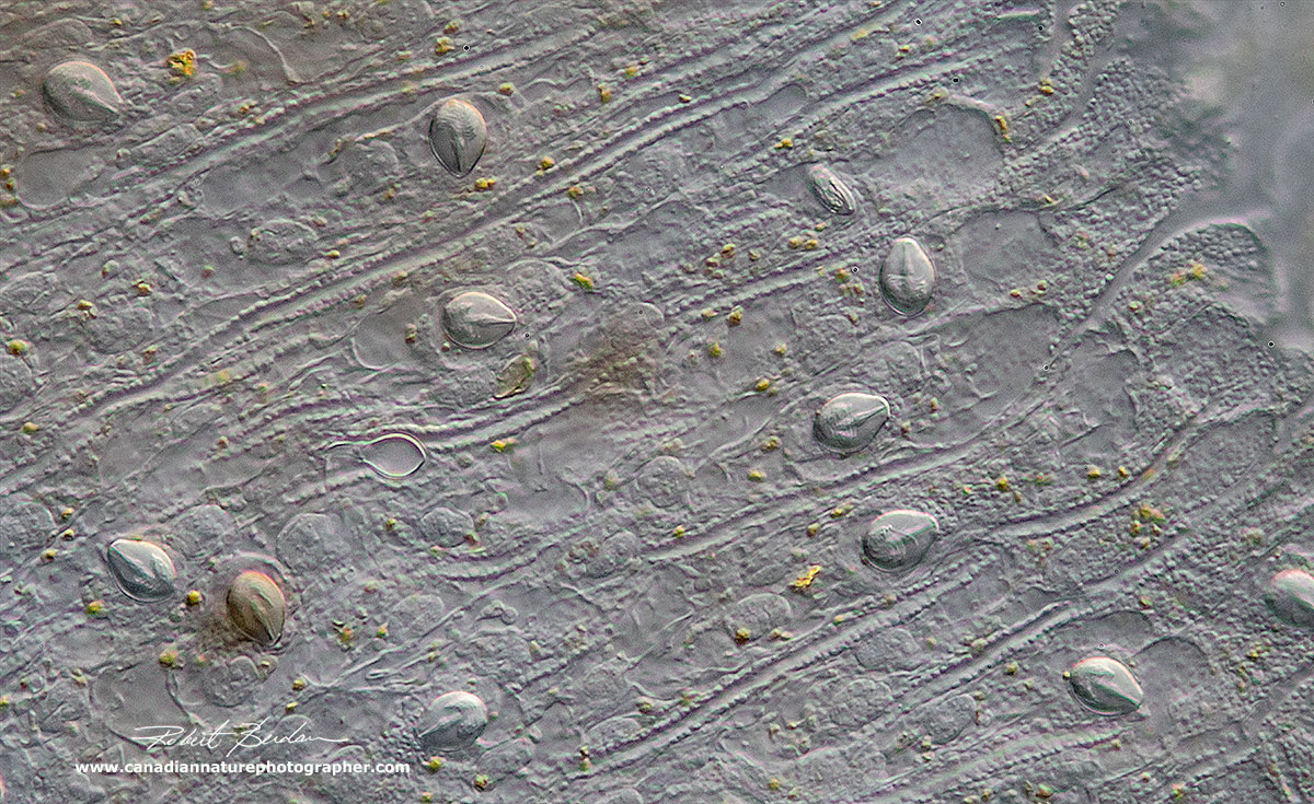
Cnidoblasts cover the Hydra's body but appear less frequently then they do on the tentacles. DIC microscopy.
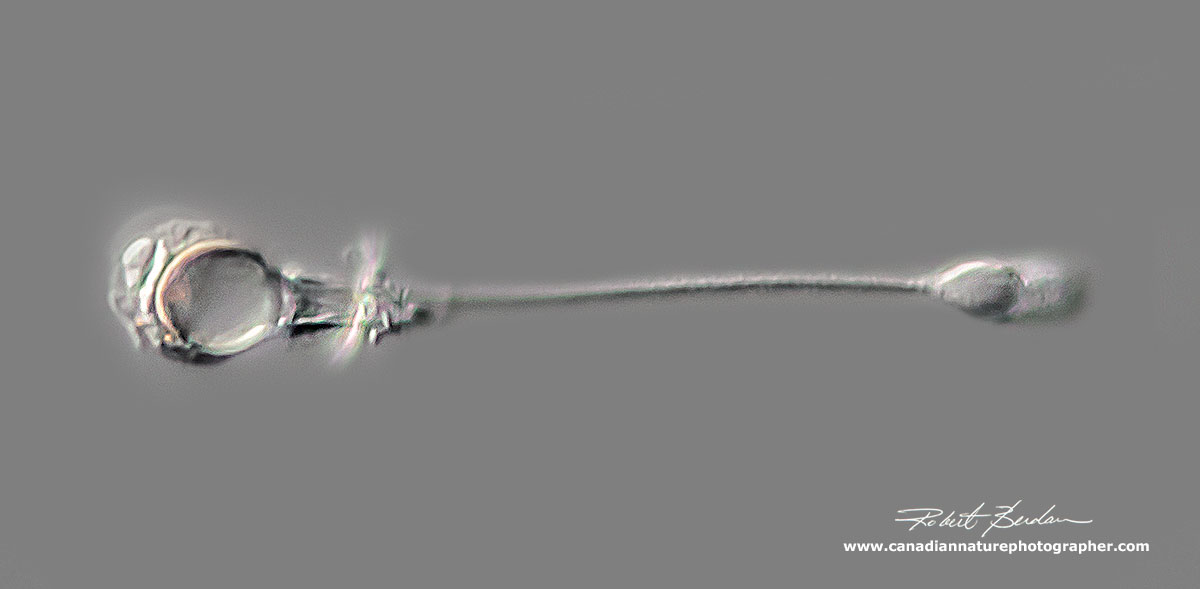
Exploded Stenotele cnidoblast from Hydra DIC microscopy

Exploded cnidoblast from Hydra DIC microscopy (not a Stenotele, but not sure which of the other three this one might be).

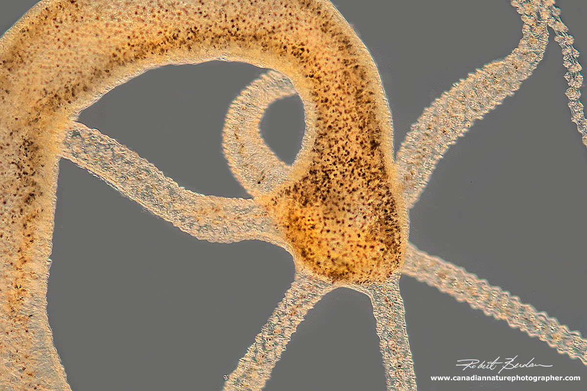
Hydra with a copepod (Cyclops) being digested inside the gastric cavity. Hydra feed mainly on Daphnia and copepods. Another crustacean about the same size as copepods called Ostracods seem to be immune to the Hydra's nematocytes and thrived in my dishes with Hydra while Daphnia and copepods seem to be the preferred food. Below is a series of photos showing a Hydra injesting a Daphnia sp.
Hydra engulfing a Cladoceran - Simocephalus sp that is much wider than its body column.


Above is a series of pictures in darkfield and bright field microscopy showing one of the brown hydras injesting a large Simocephalus sp. The mouth is able to expand much wider then the Hydra's body column in order to fit the entire animal.

Above shows a photo where I brought a damaged Simocephalus close to the hypostome of a brown Hydra (dotted lines show the hypostome opening) and I watched the hypostome expand as the tentacles grabed hold of the Daphnia. I wondered how the Hydra is able to swallow something that was much wider in diameter that its own body. When the mouth is closed it is a continous sheet sealed with septate junctions (these junctions are unique to invertebrates). The initiation point of mouth opening is defined by a morphologically distinct group of cells at the tip of the hypostome and the mouth can open wider than the body column during feeding as shown in photos above where one is feeding on Daphnia.

Above shows a closeup of the expanded hypostome using DIC microscopy. The arrows show the outline or opening of the hypostome. Recent research indicates that the hydra must tear through its epitheilial tissue each time it opens its mouth and the opening is controlled by radial myonemes (muscle like cells) in both the endoderm and ectoderm (Carter et al. 2016). Also see brief article about How Hydra Eats (A.R Dunn 2016) for why understanding this simple process is valuable in understanding complex tissue movement in general.

Hydra viridissima colour inverted to show the green algae (Chorlella) more clearly
Taxonomy of Hydra
Kingdom: Animalia
Phylum: Cnidaria
Class: Hydrozoa
Order: Anthoathecata
Family: Hydridae
Genus: Hydra
Regeneration and Aging Studies
Much of Hydras ability to regenerate parts of its body is due to stem cells. Hydra can even be dissociated into single cells and the cells will reform aggregates and regenerate. In 1998 a study by D. Martinez suggested that some Hydra do not age and this has led to new studies trying to uncover the mechanism by which this animal is able to do this. Among 259 human aging genes 207 (80%) were conserved in Hydra oligactis (Tomczyk et al 2014). The aging-induced regulation of these genes is currently under investigation. One gene (Fox0) which regulates in response to stress affects the proliferations of stem cells in some organisms but knocking out this gene does not seem to have a deleterious affect in Hydra. One species of Hydra oligactis can be induced to age by transferring it to cold water and this organism shows promise in regards to studying the genes involved in aging. While most of us might simply watch and photograph these amazing organisms because they are common in ponds remember that studying even small organisms might lead to important discoveries.
Photomicrography
All photographs are of live Hydra and were collected from a fishing pond in the foothills. For details on how I took these pictures please see my article - Tips on How to Take Better Pictures with a Microscope - Photomicrography. In brief, I used a Nikon D500 camera ISO 400, Zeiss Axioscope with DIC, Bright field, and Darkfield lighting and controlled my cameras with free software Digicam control. I held the Hydra for photography by compression under the coverslip. Because Hydra are relatively large I often used low magnification objectives 2.5, 5 and 10X in order to capture the entire animal. Focus stacking was sometimes used to increase the depth of field in the photographs. See my article on focus stacking if you are interested in how I do this.
Summary
Hydra is an extremely interesting animal to observe with a microscope and has several features that make it suitable for studying regeneration and aging. The organism can be found in pond water and different species can be distinguished based on the presence or absence of symbiotic algae, morphology of their cnidocytes and the pattern of tentacle branching on new polyps. Hydra can also be purchased for study in classrooms from Biological supply stores. They can be cultured and future studies into the genetics and molecular biology of this organism may lead to a better understanding of how humans age and may suggest ways to extend our lives. RB
Links and References
(2018) Hydra - Wikipedia
(2016) Carter. J.A et al. Dynamics of Mouth Opening in Hydra. Biophysical Journal 110: 1191-1201 - download PDF.
(2016) A.R. Dunn. How Hydra Eats. New and Notable Biophysical Journal - download PDF
(2015) M. Simon Absurd Creature of the Week: This amazing little critter just might be immortal. www.wired.com
(2014) S. Tomcyzky et al. Hydra, a powerful model for aging studies. Invertebrate Reproduction & Dvelopment. 59: 11-16 - download PDF
(2012) Investigating the Agelessness of Hydra - online blog.
(2012) M. I. Deserti and M.O. Zamponi. Biometric and statistical investigations on the cnidoma of the genus Hydra (Cnidaria, Hydrozoa). Iheringia, Serie Zoologia, Porto Alegre, 102: 298-302 - download PDF.
(2010) J.S. Hwang et al. Nematogalectin, a nematocyst protein with GLyXY and galectin domains, demonstrates nematocyte-specific alternative splicing in Hydra. PNAS 107: 18539-18544 - download research PDF
(2002) G. Kass-Simon and A.A. Scappaticci The behavorial and developmental physiology of nematocysts. Can. J. Zool. 80:1772-1794 - download PDF
(2002) G.O. Mackie What's new in cnidarian biology. Can. J. Zool. 80: 1649- 1653 download PDF
(2001) S. Szczepanek, M. Cikala and C.N. David. Poly-Y-glutamate synthesis during formation of nematocyst capsules in Hydra. J. Cell Science - research paper download PDF
(1998) D. E Martínez. Mortality Patterns Suggest Lack of Senescence in Hydra. Experimental Gerontology. 33:217-225 - Download PDF
(1983) Hydra Research Methods H.M. Lenhoff (ed.) Plenum Press. N.Y. 413 pp. Includes keys and methods for handling and photographing Hydra for research purposes.
(1983) R.D. Campbell. Identifying Hydra species. In: Lenoff, H.M. ed Hydra: Research Methods. NY, Plenum Press. pp 19-28.
(1959) L. H. Hyman Coelenterata in Fresh Water Water Biology 2nd ed W. T. Edmondson ed
John Wiley, NY pp 313-322 contains keys to Hydra view online book here.
(1953) W.J. Garnett. Freshwater Microscopy. Constable & Co. Ltd. London. pp 175-184. Although an older book the black and white photomicrographs are excellent and the text is easy to read -used books are available from ABE books in England.
Authors Biography & Contact Information

Robert Berdan is a professional nature photographer living in Calgary, AB specializing in nature, wildlife and science photography. Robert retired from Cell\Neurobiology research to take up photography full time years ago. Robert offers photo guiding and private instruction in all aspects of nature photography and Adobe Photoshop training - including photomicrography, macrophotography.
Related Microscopy Articles by Robert Berdan on this web site
1. Photographing Daphnia
2. Photographing Gastrotrichs
3. Photographing Rotifers
4. Photographing Ciliates
5. Photographing Stentors - A Large Unicellular Protozoan (ciliate) living in Freshwater
6. How to Collect and Photograph Water Bears (Tardigrades).
7. Tips on How to take Better pictures with a Microscope
8. Microscopic Pond Organisms from Silver Springs Calgary
9. Microscopic Life in Ponds and Rainwater - Pond Scum I
10. Photographing Microscopic Plant and Animal Life - Pond Scum II
11. Photomicrography and Video of Protozoa, Volvox and Rotifers
12. Home Microscopy Laboratory for Photomicrography
13. The Art & Science of Photomicrography with Polarized Light
14. Photographing Through a Microscope Photomicrography - Inner Space
15. Focus Stacking comparing Photoshop, Helicon Focus and Zerene
16. Rheinberg Filters for Photomicrography
17. Scanning Electron Microscopy - Photography
18. Photomicrographs of Diatoms from 1877 by John T. Redmayne
Email at: rberdan@scienceandart.org
Web site: www.canadiannaturephotographer.com
Phone: MST 9am -7 pm (403) 247-2457.
Click on the buttons below and share this site with your friends