
How to Collect and Photograph Water Bears (Tardigrades)
Dr. Robert Berdan
June 14, 2018 (updated July 3)
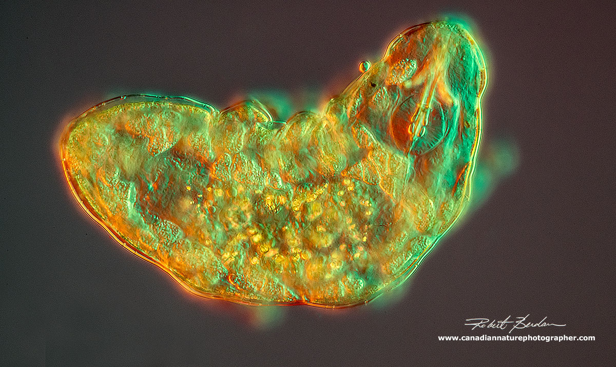
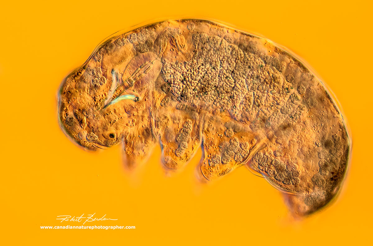
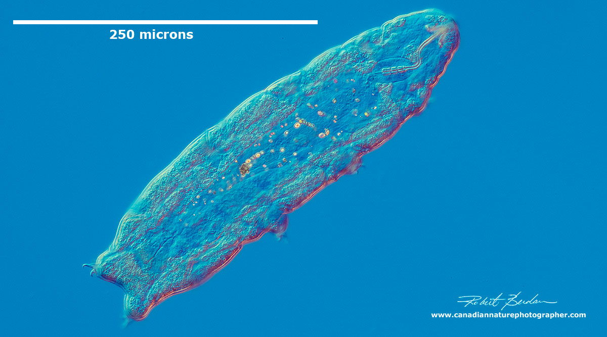
Golden water bear viewed by a combination of DIC (Differential Interference Contrast Microscopy) and Rheinberg Lighting 400X.
Introduction
As a biologist and photographer I am drawn to the beauty that is all around us; sometimes it's big and sometimes it's small. By looking and learning more about some of the smaller organisms I am amazed at what I see. It's my hope to share this hidden world with you so that you might become curious and look for it, read about it, or simply look more carefully the next time you are outside. It doesn't matter if you live in a big city, are near a park, have a backyard, or visit the wilderness - water bears live are all around us. Recently a new species of water bear was discovered in a parking lot in Japan. You might even be inspired to become an amateur scientist.
In this article I will share some of my recent pictures of "water bears". They are also called "moss piglets" and belong to the Phylum Tardigrada which means "slow walkers'. These tiny animals range in size from 50-1200 microns (micron = 1\1000 of a millimetre) - basically they are about 0.5 mm in size and they can be seen with your naked eye as a small white spot. When magnified they can be seen to hobble along on their eight legs. Most Tardigrades are white or translucent and have a world wide distribution. So far about 1200 species have been described and it's likely more will be discovered. They are related to the arthropods.

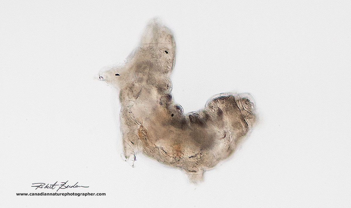
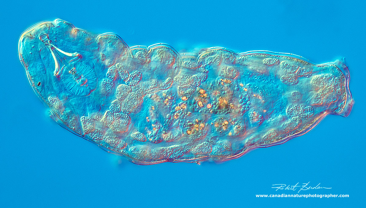
Water bear (Tardigrade) Dorsal view 200X DIC microscopy - note eyes on the left
Water bears live in the interstitial spaces of moss, lichen, liverworts, soil and leaf litter where they are surrounded by a thin film of water. When the water dries up they shrink to form cysts or tuns that can survive extreme temperatures, UV light, and low oxygen. One of the reasons there is scientific interest in these small animals is because we don't completely understand how they are able to survive these extremes. Most cells when frozen are killed by water crystals that form, water bears are able to suppress the ice crystal formation by displacing the water with sugars.
Some water bears are found in water, but most are limnoterrestrial, that is they depend on water drops that adhere to moss or other plants. They have life spans of about 3-4 months, but when they become a cyst or tun they can survive in this state for years. Studies also suggest that water bears show little specificity for specific species of moss or lichen.

Moss and lichen - ideal habitat for Water bears
Water bears feed using stylets that pierce the plants and they suck out the food. Some Tardigrades prey on other water bears, nematodes, protozoa and rotifers (see my previous article to see pictures of these microorganisms). Star Trek fans might have noticed that the new show features a giant water bear they feed pollen which in turn allows the spaceship to jump across space - far fetched but it's all fantasy anyway. Tardigrades, however have been shown to survive the vacuum of open space and solar radiation for at least 10 days which is another reason scientists are interested in them. However many biologists study them simply because they are fascinating - let's have a look.

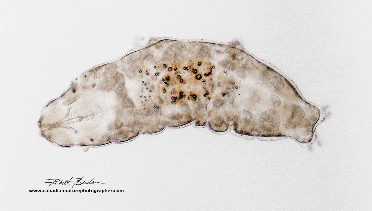
Water bear from the side 200X DIC microscopy. Inside the animal you see the individual cells, the large cells at the top are oocytes (eggs). Below the body there are three paired limbs and on the posterior end are two more limbs. Each limb usually has distinctive claws which are not visible in this photo but see below.
Where to find Water bears
Water bears are found almost every where there is water - including the Antarctic. The first place to search for water bears is in moss or lichen - miniature forests. I collected some moss next to the Silver springs waterfall and brought the moss home in a paper bag. I soaked the moss with distilled water and next day found several Tardigrades swimming in the dish. I used a stereomicroscope to find them but you could use a magnifying lens. They are not the only thing living in the moss, you will see other things moving around; most commonly nematodes, ciliates, rotifers, mites, and springtails (Collembola). I am now collecting a wide variety of different mosses and lichens in search for different species of water bears. This is the beginning of my quest.
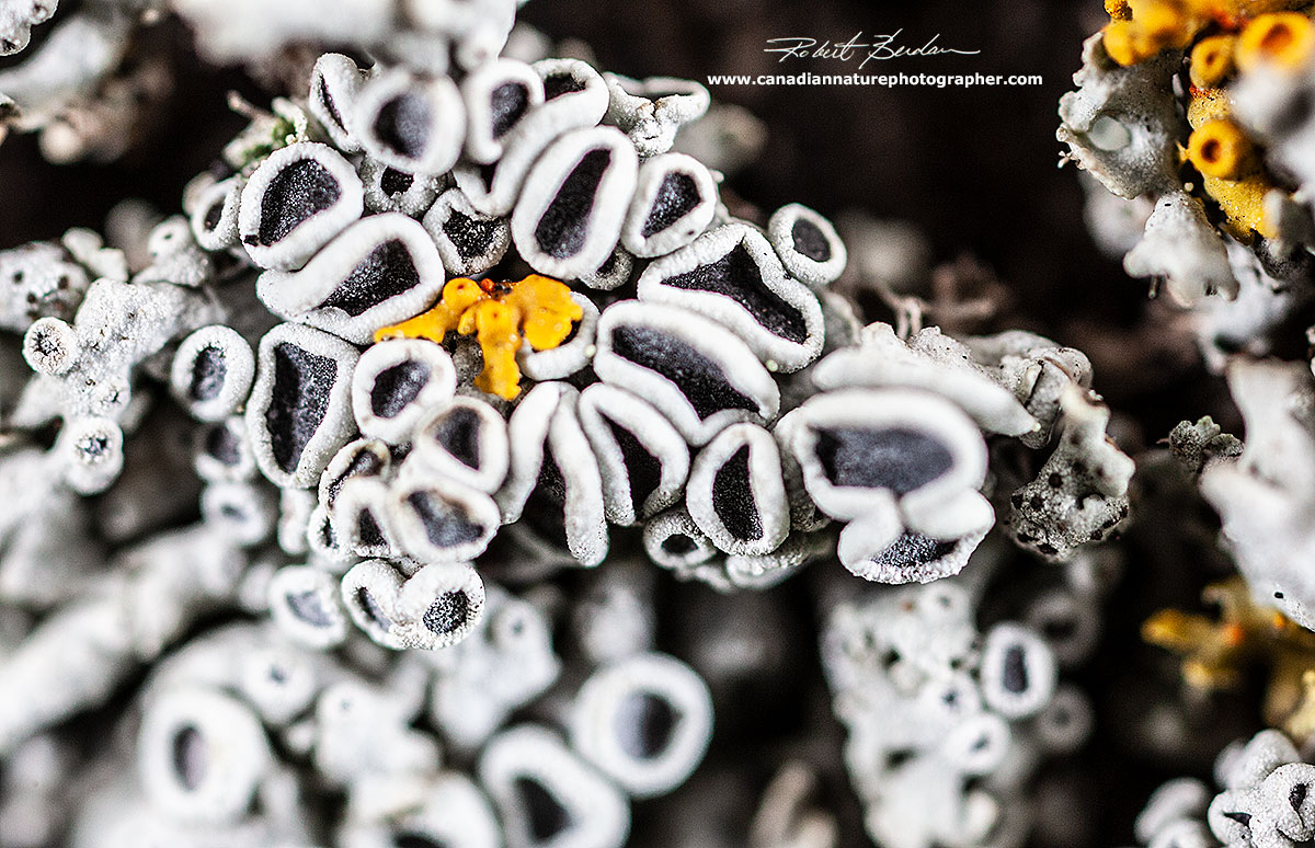
Dog tongue lichen (Peltigera aphthosa) - grows in moist places, on the ground, on trees and rocks. I found this lichen to be a rich source of water bears.

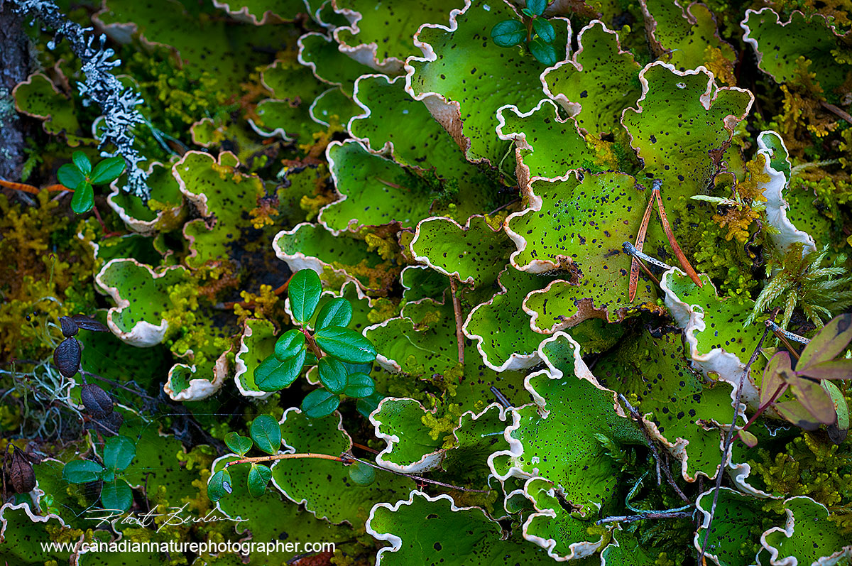
Lichen and moss growing on a rotting log in Yoho National Park. Many of the mosses, liverworts (Bryophytes) and lichens are beautiful in themselves - take notes and pictures of the places you collect the plants when looking for water-bears. Tall moss Cladonia subradiata?
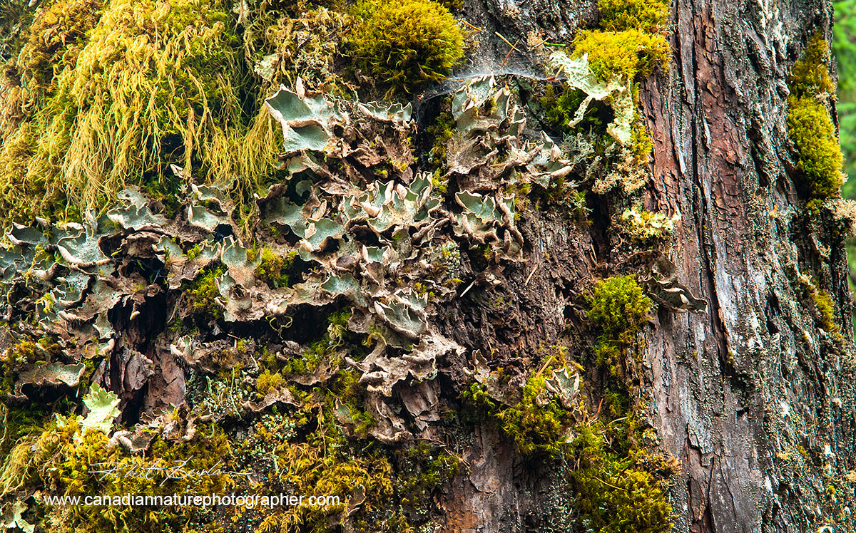
Tree from the West Coast of British Columbia showing a wide variety of lichen and moss growing on it. This would be an ideal spot to collect moss and look for water bears. Tree moss supposedly contains a greater variety of water bear species.
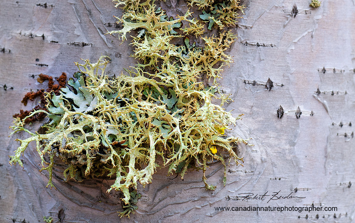
Lichen growing on a birch tree. Water bears frequent tree lichen. To extract the water bears leave the lichen in distilled or rain water over night and search the water around the moss the next day. Several species of lichen above: lime green colour - Hypogymnia physodes front - Cladonia sp., red and yellow coloured lichens - ?.
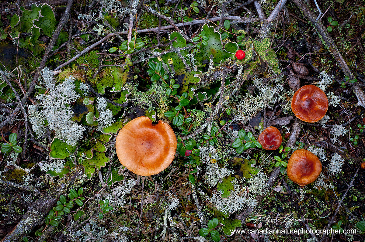
A wide variety of Lichen and moss growing on the Tundra along with mushrooms in Yellowknife, NT. I plan to collect water bears from the tundra around the end of this summer. Several species of lichen are shown: light colour Cladina rangiferina, Green - Peltigera aphthosa.
Equipment Needed to See & Photograph Water bears
Water bears occupy moss, lichen, liverworts and leaf litter. You can collect small samples of the plants and place them in paper bags - I use lunch bags from Walmart. Label the bags with information, date, location (GPS info) and other information that might be useful. You can store the samples in the paper bags for weeks - possibly longer. Once your are home you can place some of the samples in a dish with distilled water (tap water may kill the water bears). I buy distilled water at the pharmacy. Alternatively you can use tap water that has been sitting for a few days to get rid of any traces of chlorine. I have seen the water bears in my dishes as soon as an hour after soaking the moss. I find the greatest number of bears by looking at the dishes the next day. It helps to view the water bears against a black background with strong light source and a stereo microscope. A magnifying lens or enlarging loupe could also be used. Look for white barrel shaped objects with eight legs that are usually moving. Some folks squeeze the water out of the moss and then examine the water. I just move the moss around in the dish and hunt for the bears. Once I find one I use a glass pipette to place them in another dish or on a microscope slide, add a cover slip, and then view them in my microscope. To preserve or fix the water bears I place them in 99% Isopropyl alcohol in a small vial and in the future I am going to start making permanent microscope slides of the bears by embedding them in Canada Balsam.
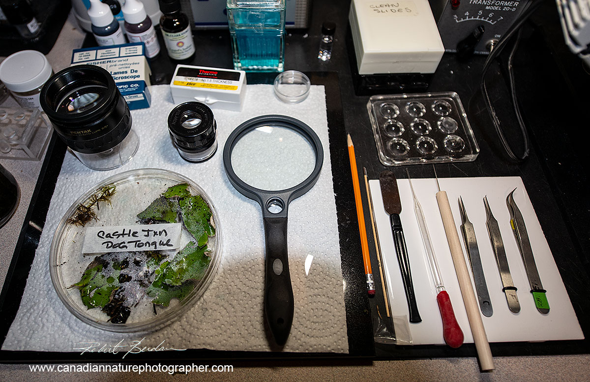
Above photo shows some Lichen in a large Petri dish, a magnifying glass and several magnifying loupes, some dissection instruments, a glass pipette, microscope slides and coverslips. All of these tools are available on E-bay and Amazon for only a small sum of money.

My work area consists of a Stereo microscope, some slides, dissection instruments and large Petri dishes with moss and lichen soaked in distilled water. Once I find the bears I transfer them to microscope slides for photography.

This is another microscopy setup that I use including an Olympus Zoom Stereo microscope and Olympus E Light microscope. Both scopes are decades old but are excellent for examination of microorganisms and photomicrography.
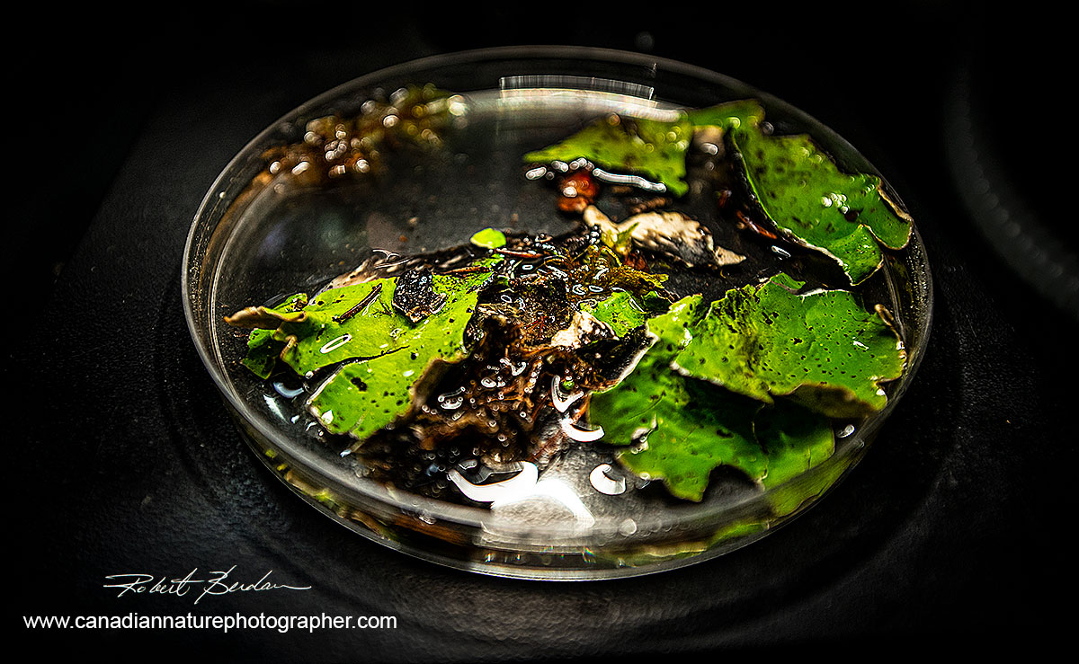
Large Petri dish with Dog Tongue Lichen (Peltigera aphthosa) soaked in water. I rummage through the dish and look for water bears.

My darkroom converted to a microscopy lab for photomicrography. I use several microscopes and stereo microscopes for photomicrography. I connect my cameras to my laptop to capture the images using free software Digicam control. A decent microscope can be purchased for a few hundred dollars e.g. AM Scopes or you can buy a used scope from Kijjii, E-bay or government surplus auctions, though it helps to learn about microscopes before you invest in a used one. My main microscope for photomicrography is a Zeiss Axioscope with DIC (Differential Interference Contrast) - white scope right side of the picture. I connected a "gaming" USB foot pedal to the laptop so I can trigger the camera and have both my hands free to focus and move the stage around.
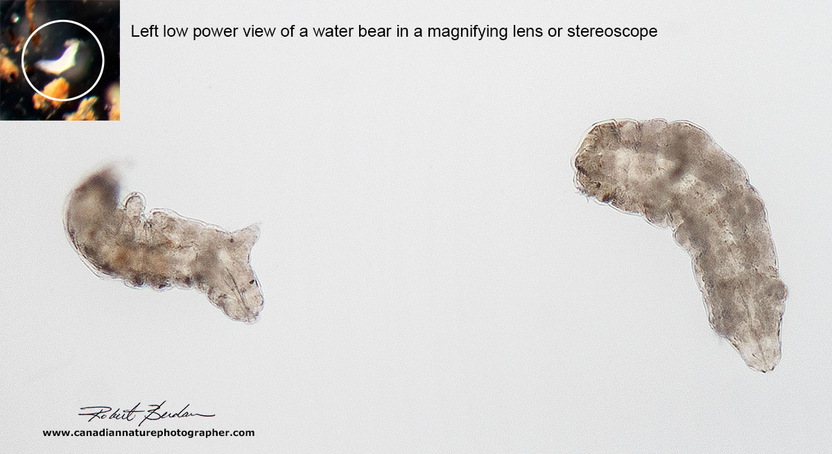
Above are two water bears as they appear in Bright field microscope at about 50X. No special lighting is required to see them. The water bear in the inset is blurred due to its movement and relatively long exposure (1\8 sec). If the water bears are moving actively I recommend you use 1\250 of sec. exposure or faster if possible; increase your camera ISO speed if necessary to achieve a faster shutter speed. I record RAW files in order to adjust the white balance afterwards so my backgrounds are white and not yellow from tungsten light bulb.
Dancing Water bear 200X Bright field microscopy. Because water bears like to move around I find it helpful to compress them using a coverslip by sucking some water out from under the coverslip with a paper towel - this slows and holds them down - see my previous article on Tips on Microscopy.
Water bear via bright field microscopy, dorsal view - 200X; see two pair of eyes on the left and the buccal pharyngeal apparatus inside the head, a higher magnification view of the head is shown below.

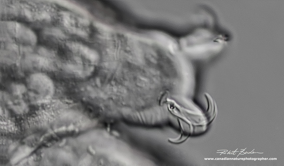
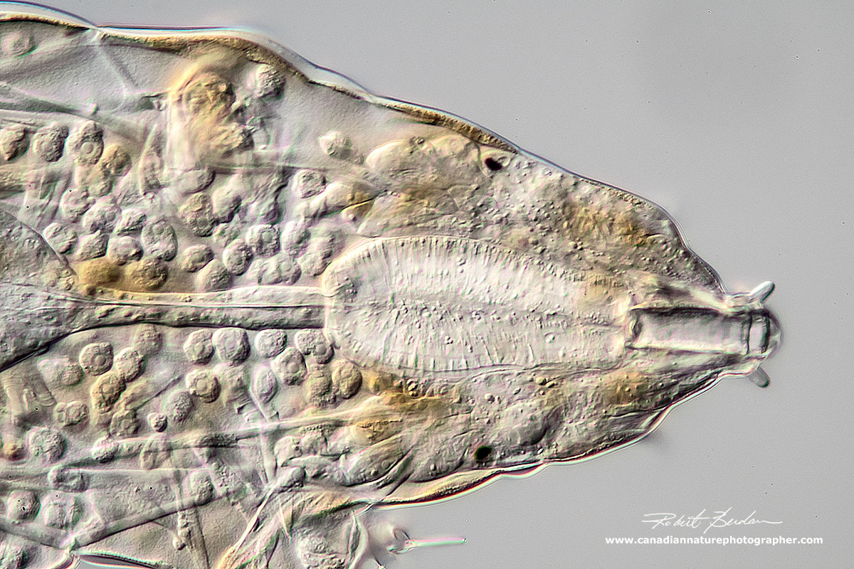
A closeup view of the head of a water bear using Bright field microscopy 400X. Note the two dark eyes on each side of the head and the buccal-pharyngeal basket (oval shaped object). See diagram below where I identify the various structures. The buccal-pharyngeal apparatus is used taxonomically to identify some species. This appears to be a member of the Macrobiotus group.
Macrobiotus hufelandi
Macrobiotus hufelandi is the most common Tardigrade typically ranging in size from 350 to 450 microns in length. The animals have Y shaped claws. The Pharyngeal bulb has two macroplacoids the first longer then the second and a microplacoid and eyespots are usually present. The hufelandi group consist of a number of morphologically similar species so without viewing their eggs its impossible to be sure if this is M. hufelandi species. Eggs are deposited free with ornamentation resembling inverted egg cups or goblets (Kinchin 1964).
Macrobiotus hufelandi (or member of this group) viewed by Phase contrast microscopy 200X. Using phase contrast it is often easier to see the shape of the claws.
Closeup of the head and Buccal-Pharyngeal apparatus viewed by phase contrast microscopy 400X. Nikon D300 on Olympus E-microscope.
Macrobiotus hufelandi viewed by DIC microscopy appears translucent, the stylets and eyes are clearly visible in this specimen.

Macrobiotus hufelandi viewed by Polarized light microscopy shows the muscle pattern and birefringent Pharyngeal apparatus and two black eyes. 200X.

Macrobiotus is also predatory - shown above feeding on a nematode. DIC microscopy
Darkfield microscopy provides a narrow cone of light to illuminate the specimen against a dark background. To produce this type of light there are special microscope condensers, or you can simulate Darkfield by using filters with a dark spot in the center, even a coin placed on a filter will work though you have to choose the right diameter coin or move the filter vertically in the light path to get the best effect. Creating the darkfield effect is easy to do on most microscopes.

Water bear 200X Darkfield microscopy -Zeiss Axioscope

Water bear 200X Darkfield microscopy, Zeiss Axioscope. The claws are out of focus, but you can see the limbs and one eye on the left side.

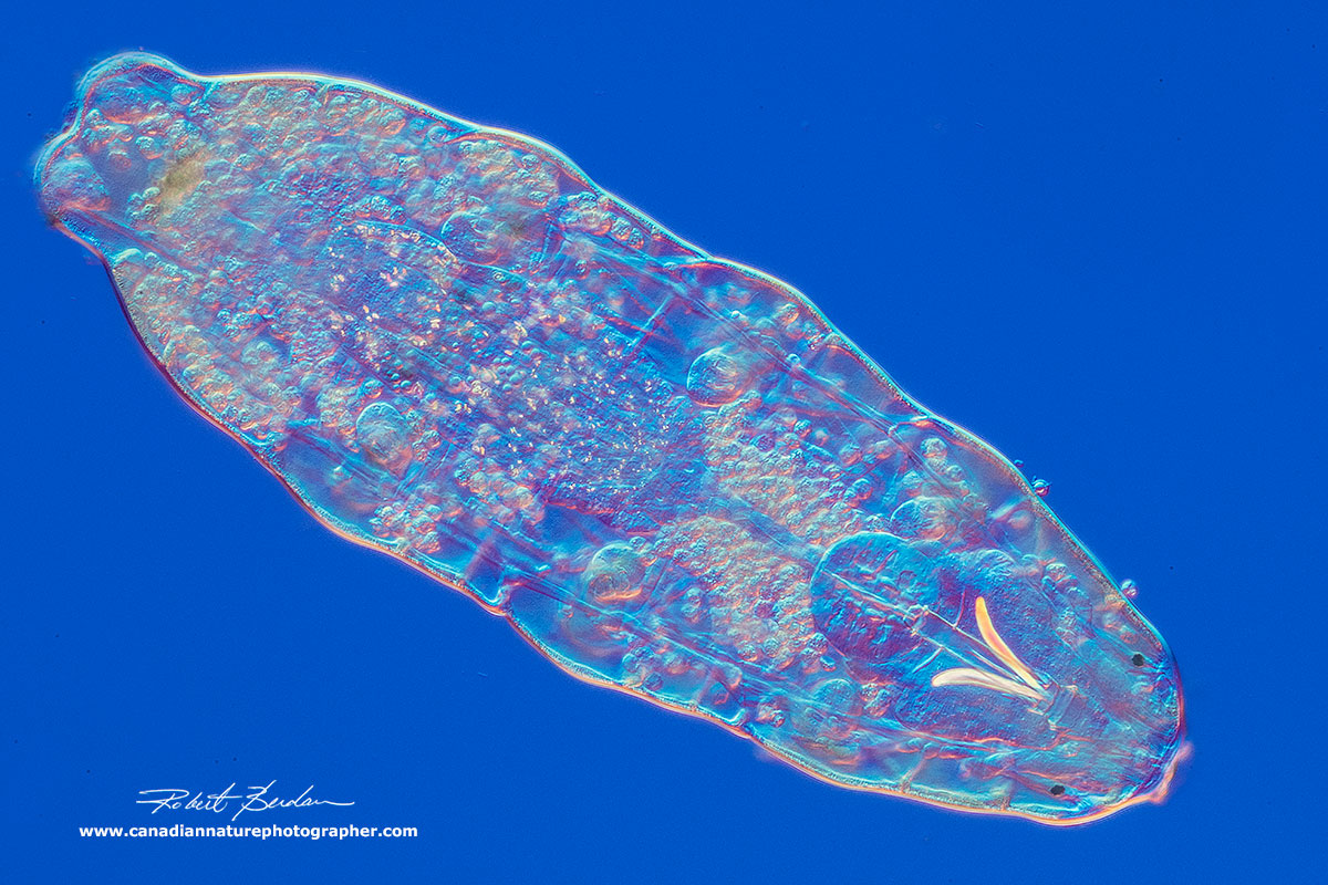
Low power view of a water bear from above. 100X DIC microscopy.
Water bear Tardigrade - DIC microscopy 200X
Water bear photographed at 200X DIC microscopy. Note the cells inside.
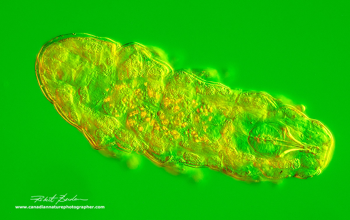
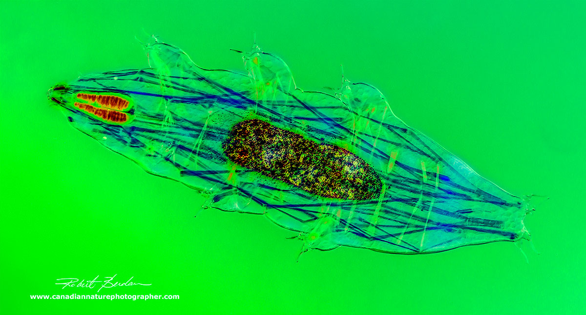
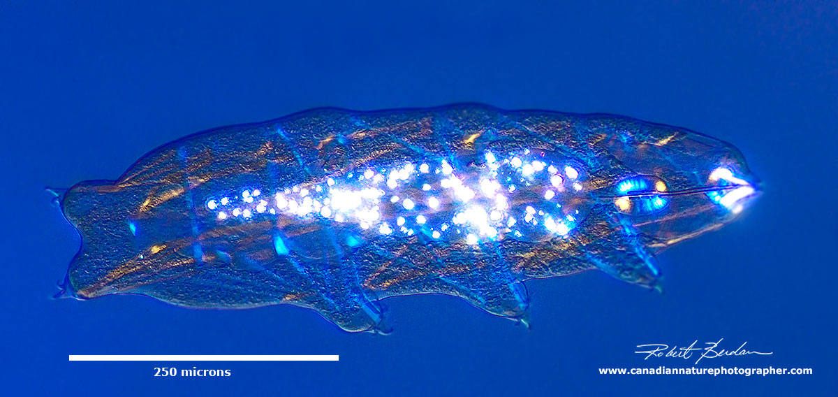
Combination of Rheinberg lighting and DIC microscopy 200X - this lighting provides yellow highlights and a green background. Rheinberg filters are available on Ebay for about $30 and can be used with almost any microscope.
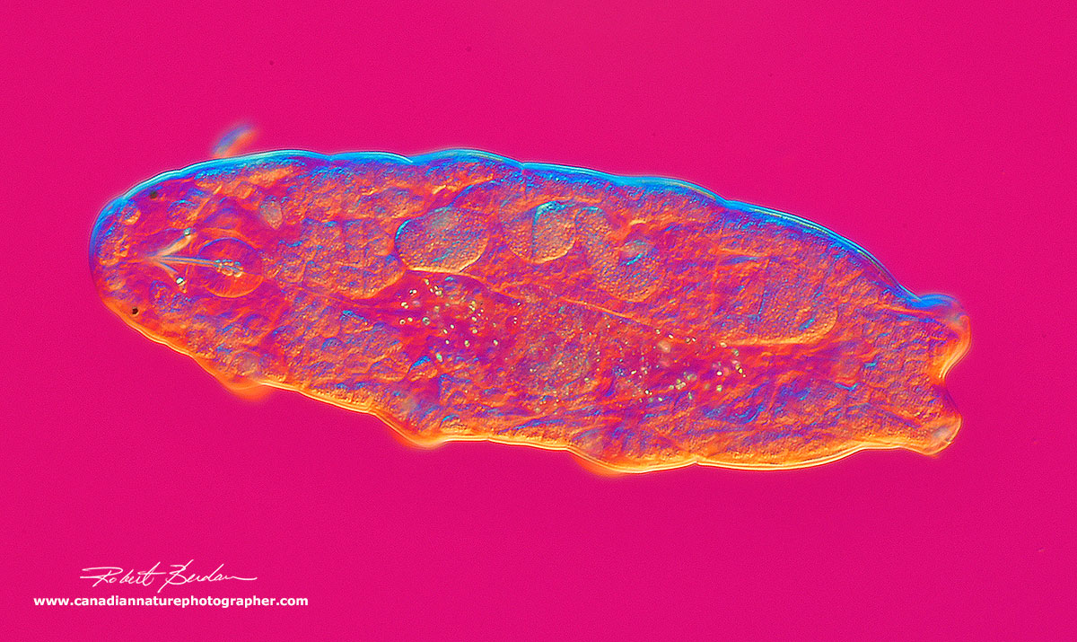
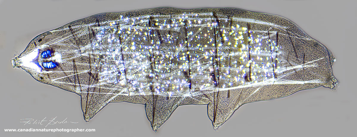
Water bear Dorsal view using DIC microscopy 200X. DIC microscopy lets me change the background colours and see the organism in a 3D relief, unfortunately DIC is expensive.
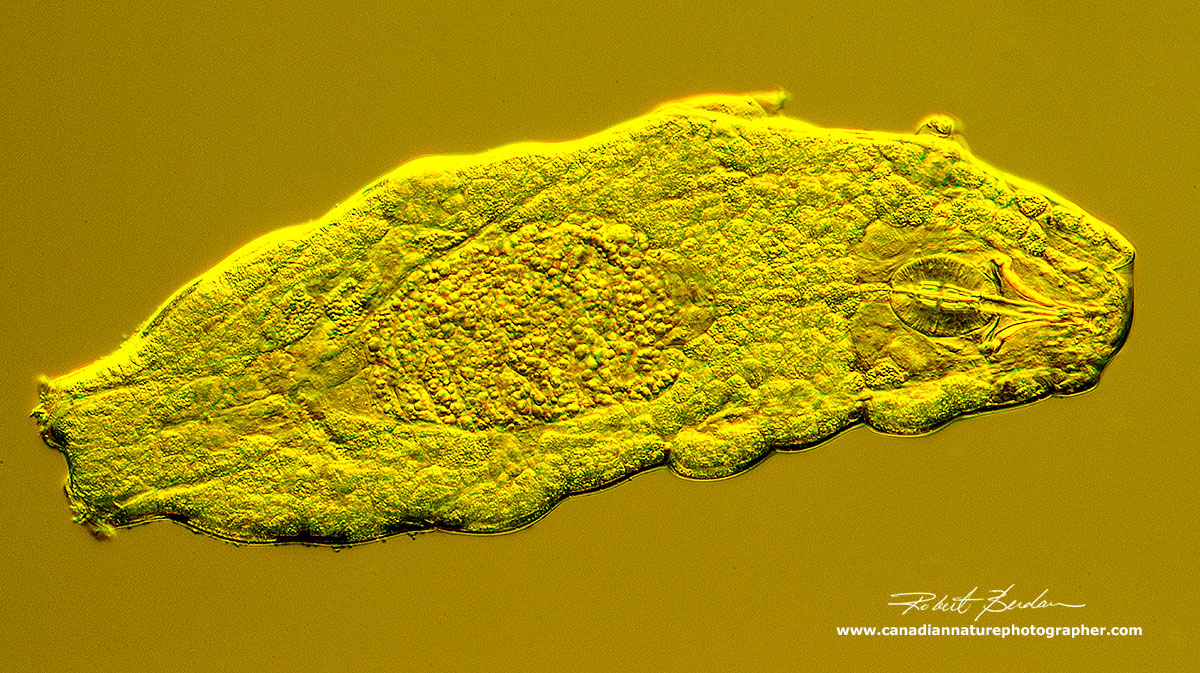
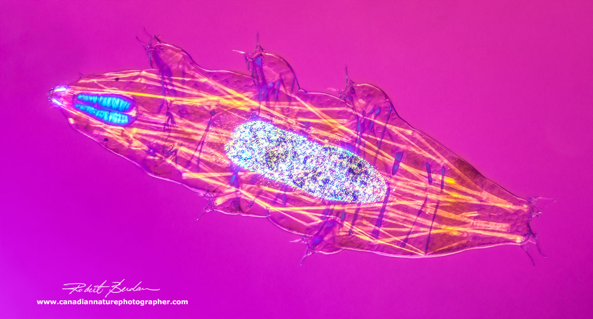
Combination DIC and Rheinberg lighting of Water bear 200X produces a golden colour.

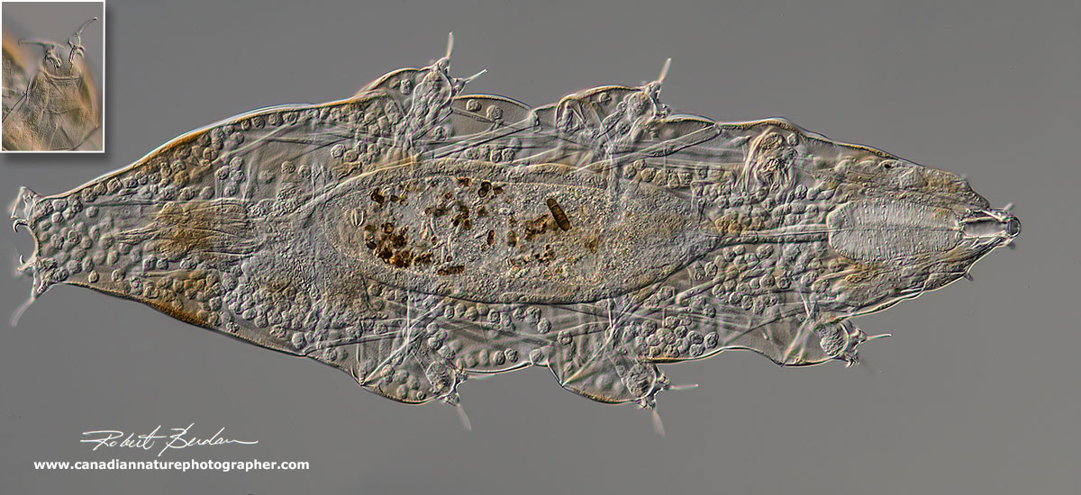
This water bear was found in Dog Tongue lichen and has some brown pigment. 200X DIC microscopy. Note the claws shown below at higher magnification. Based on the fact this Tardigrade has a long primary claw, round pharynx, 2 macroplacoids, thin buccal tube and brown pigment stripes it is likely Ramazzottius oberhaeuseri (as suggested by Dr. Gary Grothman).

Close up Ramazzottius oberhaeuseri showing the claws - 600X DIC microscopy There are basically Symmetric 2 1 1 2 and Asymmetric claw patterns 2 1 1 2 where 2 represents a small secondary branch and 1 represents a large primary claw.

Eutardigrade claw shapes from: Phylogeny of Eutardigrada: R. Bertolani et. al. (2014) Molecular Phylogenetics and Evolution 76: 110-126.

High magnification photo showing claws on the end of the legs. 600X DIC microscopy.
Y shaped Tardigrade claws characteristic of Macrobiotus sp (C above) 1000X DIC

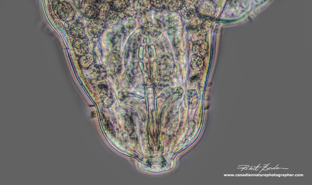
High magnification picture showing the Buccal - Pharyngeal bulb, 2 stylets, 3 sets of macroplacoids and 2 smaller microplacoids DIC microscopy 800X.

Buccal-Pharyngeal apparatus - showing the piercing stylets and some coelomocytes - round cells inside the Tardigrade. The Pharyngeal apparatus is important taxonomically, along with the claws and cuticular structure.
Polarized Light Microscopy of Water Bears
Polarized light microscopy is often used to show birefringence - the ability of some crystals and anistropic substances to allow polarized light to pass through crossed polarizing filters. In animals muscles have a regular arrangment of fibers and molecules and show birefringence. By adding a wave plate between the polarized light I am able to change the background colours just as I am with DIC microscopy. In the photos below you can see the muscle arrangement - for instance in the bottom photo the muscles are blue and whereas in the middle photo they are white. The buccal-pharyngeal apparatus is also birefringent as well as various intracellular organelles.
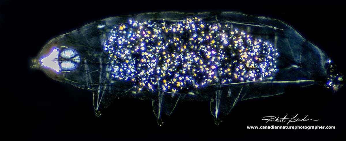
 Water bears view via Polarized light microscopy 200X. These photos show the muscle arrangement in the animals.
All the Tardigrades in this article belong to the Class Eutardigrada. I have identified them using various keys, but apparently to identify some species it is also necessary to look at the structure of their eggs. Many species are similar and correct identification often requires the assistance of experts who have worked with Tardigrades. All the Tardigrades shown in this article are the most common species encountered.
Predatory Rotifer - Millenisum tardagradum
I collected lichen off a dead pine tree branch and soaked it with distilled water and found what I believe is Millenisum tardagradum - a predatory Tardigrade based on its size, lack of placoids on the buccal pharyngeal appartus, 2 pairs of papilla on its head around the mouth and claw shape. Inside the gut I can see a rotifer mastax - proof this is a predatory tardigrade.
Milenisium tardigradum has a large elongated muscular pharynx and inside the midgut a rotifer mastax can be seen. Milenisium is known to prey on rotifers. The Tardigrade was 500 microns long (0.5 mm) and was found in water soaked with Lichen Hypogmnia physodes (or closely related species of lichen) collected in the rocky mountains. The inset shows an enlargment of the Tardigrade claw structure, and unlike the other Tardigrades I have been photographing this one looks fierce. Photographed with a 20X objective and photos stitched together.
The structure of the pharyngeal bulb of the apochelate Milnesium tardigradum is elongated and placoids are absent. There are two oral papilla one pair is out of focus but you can see them in the lower magnification photo above. Class: Eutardigrada; Order: Apochela; Family: Milnesiidae.

Above are two photographs are of Milenisium tardagradum photographed with polarized light microscopy showing the muscle arrangements. Birefringent material inside the midgut appear granular and the muscles in the buccal pharngeal apparatus appear bright blue - 200X Polarizing microscopy.
Diphascon (Hypibiidae Family)
Diphascon scoticum (or group?) has been widely recorded and the animals are usually between 200-370 microns long. Thin macroplacoids are present, usually increasing in length from first to third (Kinchin 1994). The stylets are shorter and the buccal tube is flexible as shown below and in this specimen the eyespots were lacking.
Diphascon species of Tardigrade DIC microscopy

Enlargement of the Buccal Pharyngeal apparatus showing 3 macroplacoids and flexible buccal tube. Also note the stylets are shoter and furtheer away from the Pharyngeal bulb. DIC microscopy.
Diphascon species viewed by Polarized light microscopy.
Water Bears in My Backyard
I have been finding water bears in almost all the lichen and moss samples I have looked at so I decided to scrape some lichen off a Mountain ash tree in my backyard and to my surprise I found water bears that appear to be Ramazzottius oberhaeuseri. The interesting thing is that I found three of them with protozoa attached to their posterior end. According to Kinchin (1994) these ciliates are Pyxidium tardigradum (Van de Land 1964). These protozoa feed on bacteria and are not thought to be directly harmful to the animal but do place a drag on the Tardigrades.
Lichen growing in my backyard on Mountain Ash tree 10X. I scraped the lichen and placed it in a Petri dish with distilled water and found several Tardigrades a few hours later. Possibly Rhizoplaca sp of Lichen.
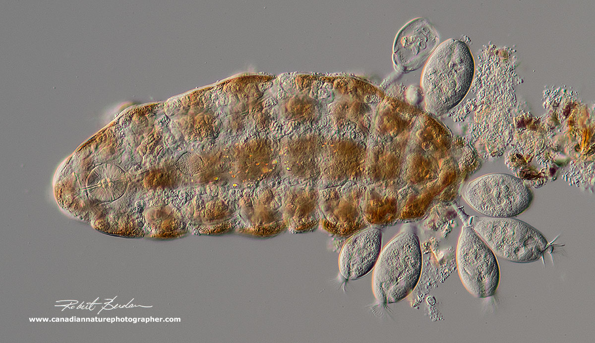
Ramazzottius oberhaeuseri with attached protozoa Pyxidium tardigradum ciliates

Closeup of the Protozoa Pyxidium tardigradum showing ciliates attached to the Tardigrade cuticle

Closeup of the Protozoa Pyxidium tardigradum showing ciliates attached to the Tardigrade cuticle. I saw one or two ciliates attached to the anterior end of some Tardigrades though most attached to the posterior end of the Tardigrade. I counted up to 9 ciliates on one animal. Reports indicate this Ciliate species is specific to Tardigrades, however I also saw them attached to a large bdelloid rotifer in the same lichen sample - see below.

Rotifer with Protozoa Pyxidium tardigradum attached 200X DIC microscopy.

Closeup of Ramazzottius oberhaeuseri Pharyngeal-buccal apparatus DIC microscopy 600X, the macroplacoids are easy to see near the center of the pharyngeal bulb in the head.
Tardigrade Tun Formation - Cryptobiosis
When limno-terrestrial tardigrades face a drastic change in their environment that can trigger them to go into a dormant state where they suspend their metabolism and are able to withstand extreme temperatures, low oxygen, absence of water, UV light, X-rays even the vacuum of outerspace. I have seen drawings and scanning electron microscope photos of tuns e.g. in the book by Kinchin (1994) but I have never seen any light micrographs of them so wasn't really sure what to look for. Dr. Gary Grothman suggested inducing the Tardigrades to form a tun. So I placed several animals on microscope slides in a drop of water, placed the slides in a large Petri dish with a wet paper towel underneath and allowed the water to evaporate slowly over two days. Sure enough I found several animals had formed tuns - see picture below (I believe the Tardigrades were from the Macrobiotus group).

Tardigrade Tun induced by Dessication on a microscope slide. DIC microscopy
Tardigrade Movie - Dancing Bears
Or watch Video on YouTube https://youtu.be/riDgQarL44A - and you can also view full screen.
References & Links
I. A. Kinchin (1994) The Biology of the Tardigrades. Portland Press, Great Britain. I got my copy on Ebay.
D. R. Nelson, R. Guidetti and L. Rebecchi (2016) Phylum Tardigrada in Ecology and General Biology Thorp and Covich's Freshwater Invertebrates, Academic Press, NW. pp 347-380.
Janice Glime (2017) Bryophyte Ecology - see her article on this web site and download all her book chapters in PDF format at the end of the article for free. She has several excellent articles about Tardigrades.
M.J. Boekner and H.C. Proctor (2005) Water-bears from the Rocky Mountains: A First Look at Alberta's Tardigrade Fauna. The Canadian Field-Naturalist. Vol 119. pp 586-588. Available online as a free PDF - search Google.
G.T. Grothman (2010) Tardigrades of Fish Creek Provincial Park, Alberta, Canada. A Preliminary Survey. The Canadian Field Naturalist. Vol 125. pp 22-26. Available online as a free PDF.
D. Stec, K. Arakawa, L. Michalczyk (2018) An Integrative description of Macrobiotus shonaicus sp. nov. (Tardigrade: Macrobiotidae) from Japan with notes on its phylogenetic position with the hufelandi group - download PDF
Tardigrades-of-North-America-New-Jersey-Survey-Michael-Shaw-NYMS-OCT2013.pdf
World Tardigrade Database - www.marinespecies.org/tardigrada
www.tardigrades.com by Martin Mach excellent selection of photos and information.
tardigraderesearch.blogspot.com - resource about Tardigrades
Tardigrada Newsletter - excellent photos and information.
M. Shaw (2016) Tardigrade Science Project book- good book for kids.
Online Key to Tardigrades (good place to start but now out of date)
YouTube video of Tardigrade
YouTube video - The Life of Tardigrades - excellent video showing different species, eggs etc
How to find a Tardigrade - YouTube Video
Enlichenment - photos and information about lichens
Electronic Altas of the Flora of British Columbia
Zeiss - Microscopy from the very beginning - free PDF
Optical Microscopy by MW Davidson and M. Abramowitz - Excellent - free PDF
Quality Microscopes in Calgary - great source for stereo microscopes
Related Microscopy Articles by Robert Berdan on this web site
1. Tips on How to Take Better Pictures with a Microscope - Photomicrography
2. Microscopic Pond Organisms from Silver Springs Calgary
3. Microscopic Life in Ponds and Rainwater - Pond Scum I
4. Photographing Microscopic Plant and Animal Life - Pond Scum II
5. Photomicrography and Video of Protozoa, Volvox and Rotifers
6. Home Microscopy Laboratory for Photomicrography
7. The Art & Science of Photomicrography with Polarized Light
8. Photographing Through a Microscope Photomicrography - Inner Space
9. Focus Stacking comparing Photoshop, Helicon Focus and Zerene
10. Rheinberg Filters for Photomicrography
11. Scanning Electron Microscopy - Photography
12. Photomicrographs of Diatoms from 1877 by John T. Redmayne
Authors Biography & Contact Information

Robert Berdan is a professional nature photographer living in Calgary, AB specializing in nature, wildlife and science photography. Robert retired from Cell\Neurobiology research to take up photography full time years ago. Robert offers photo guiding and private instruction in all aspects of nature photography and Adobe Photoshop training - including photomicrography, macrophotography and searching for water bears!
Email at: rberdan@scienceandart.org
Web site: www.canadiannaturephotographer.com
Phone: MST 9am -7 pm (403) 247-2457.
Click on the buttons below and share this site with your friends