
Vitamin B12 and Sodium Chloride Crystals
by Light Microscopy
by Dr. Robert Berdan
March 5, 2023
Vitamin B12 crystals by polarizing microscopy. Crystals grew from the edge of the coverslip 400X.
Introduction
I was contacted by an agricultural laboratory in Texas that asked me to photograph some products they work on: pecans, cotton fibres and vitamin B12 also called Cobalamin. I was fascinated with vitamin B12 and its importance in our diet. Vitamin B12 is an essential vitamin that forms red crystals when purified and examined by light microscopy (E. Simone et al. 2015). Vitamin B12 is required for DNA, fatty acid, and amino acid synthesis. A deficiency can result in pernicious anemia, neuropathy and in tiredness, muscle weakness, dizziness, poor reflexes, confusion, pale skin and decreased appetite in people over 60. The vitamin is usually absorbed by consuming meat or animal byproducts such as eggs, milk and butter. Vegetarians and vegans are recommended to take supplements to get enough vitamin B12. Someone with a serious deficiency in vitamin B12, doctors prescribe injecting it intramuscularly into their patients (my mother required this in her 80's). If you suspect a vitamin B12 deficiency see your doctor for advice. Vitamin B12 is also used for the treatment of oral cyanide poisoning (J. Lee et al. 2016).
Vitamin B12 is made by bacteria and archaea (H. Fang et al. 2017). It can be synthesized (Wikipedia) but the process is difficult and offers small yields. In ruminants like cows and sheep which feed on plants and are foregut fermenters, microbial fermentation occurs in the rumen before entering the true stomach and provides Vitamin B12 produced by bacteria. Some mammals like rabbits, pikas and beavers that consume high fibre diets will feed on their feces. This process is called cecotrophy or coprophagy. The reason is that re-ingestion allows for the absorption of vitamin B12 that is normally produced by bacteria in these animals cecum and large intestine (hindgut fermentation). Curious readers might want to read more about why do some animals - including your dog - eat Poop? Also see (O. Soave and S.D. Brand 1991).
Vitamin B12 is a complex molecule based on a corrin ring similar to the porphyrin ring found in heme (blood) but the central ion is cobalt instead of iron.

Chemical structure of Vitamin B12 courtesy of Wikipedia commons
Below I describe methods I used to make Vitamin B12 crystals and examine them by light microscopy.
Methods & Equipment
First I had to grow crystals of vitamin B12 on microscope slides. Pure or highly purified chemicals are not easily available to the public. Sigma offers 500 mg for about $117.00.

Methylcobalamin (shown above) is a form of vitamin B12. Physically it resembles the other forms of vitamin B12, occurring as dark red crystals that freely form cherry-colored transparent solutions in water. Image from Wikiwand creative commons license photo by S.B. Harris.
I searched a local pharmacy store and picked up a bottle of pills (Jamieson's Vitamin B12 2,500mg, cost under $20.00 see below). The small pink pills were solid so I ground them up in a mortar and pestle and then dissolved them in distilled water, ethanol and a mix of water and ethanol 1:1 in volume. I placed samples on microscope slides and allowed the slides to dry at room temperature. I also heated some of the samples on a hot plate to accelerate the evaporation of the solvent as this sometimes results in the formation of crystals when using different chemicals.

Above is a bottle of vitamin B12 from the pharmacy and on the right is a 1 ml ampule from the Medicine Shop containing injectable vitamin B12 solution. Only vitamin B12 in the ampule produced crystals I could view and photograph by light microscopy.
I did not see any crystals form when using Jamieson's pills. On the Jamieson bottle they listed cellulose, dicalcium phosphate, vegetable magnesium stearate, hydroxypropyl celluloses, and silice (SiO2) - sand. I could see some cellulose fibres using a microscope, but no crystals. Simone et al. (2015) indicated that impurities significantly inhibited vitamin B12 crystallization. I tried to remove impurities by filtering the solution and centrifuging the solution so any insoluble impurities would appear at the bottom of the tube. I repeated the experiments and still no crystals formed. I tried heating the solution in a water bath at 60 °C for an hour and then placed drops on a microscope slide, but again no crystals formed. I air dried and heated some sample solutions on a microscope glass slide and still did not see any crystals.
I purchased some vitamin B12 that is used for injection into patients. The vitamin B12 was in liquid form and mixed in sterile 0.9% saline solution. The solution was bright red in colour and it was sold in 1 ml ampules for $6 each from the Medicine Shop in Calgary (see photo above). I placed small drops of the injectable vitamin B12 onto microscope slides. I heated the slides and let some slides dry at room temperature. On some slides I covered part of the drops with a coverslip. Heating did not seem to significantly promote vitamin B12 crystallization, but most of the crystals of vitamin B12 formed under coverslip. The crystals would grow inward from the edge of the coverslip. When I added some vitamin B12 powder from the Jamieson tablets to the solution on the slide to act as seeds, no additional crystals formed. I didn't see a lot of crystals, but eventually found enough to take some pictures. Vitamin B12 crystals were small, bright red and formed rectangular shaped crystals that formed in clusters. The spikes were about 50 to 200 microns long (micron = 0.001 mm) and 10-20 microns wide with lots of variability in crystal size.
I used three microscopes: Motic BA310 polarizing scope and two Zeiss Axioscopes with polarizing filters, dark-field and one equipped with differential interference contrast (DIC) and Rheinberg lighting. I used 4, 10, 20 and 40, 63X objectives. Each microscope had a 35 mm digital camera attached (Nikon D800, D500, Canon 5D Mark II) and I captured RAW files using free software (Digicam control) on a PC computer running windows. RAW files were processed in Adobe Photoshop CC2023. Colours shown were captured by the microscope techniques and not by any image manipulation.
Results
Vitamin B12 crystals growing on the edge of a coverslip by polarizing microscopy 400X.
At first I found no crystals using Jamieson's crushed pills though I could see cellulose fibers and other undissolved material. After reading a scientific manuscript (E. Simone et al. 2015) they reported that even small impurities could significantly inhibit vitamin B12 crystals, so I repeated my experiments again trying to remove contaminants from the pill solution, but this made no difference.
In contrast, samples of vitamin B12 from the ampules, dried on microscope slides at room temperature, and samples heated, provided a variety of needle-like crystals under the coverslip. Other crystals observed appeared pyramidal in shape from above and further investigation showed that these crystals were salt crystals (sodium chloride). Some of these pyramidal shaped crystals exhibited glowing colours around their edges with Differential Intereference micoroscope (DIC). Salt is not coloured or birefrigent (see photos below) by polarized light microscopy. In bright-field microscopy the salt crystals from the ampules had red colour on their surface from the vitamin B12 solution.
Vitamin B12 crystals forming on the edge of a coverslip, bright-field microscopy 400X.
Vitamin B12 crystals that formed on the edge of a coverslip, bright-field microscopy 400X.

Vitamin B12 crystals that formed under a coverslip and dried on a microscope slide, bright-field microscopy 400X This crystal was not attached to the coverslip edge.
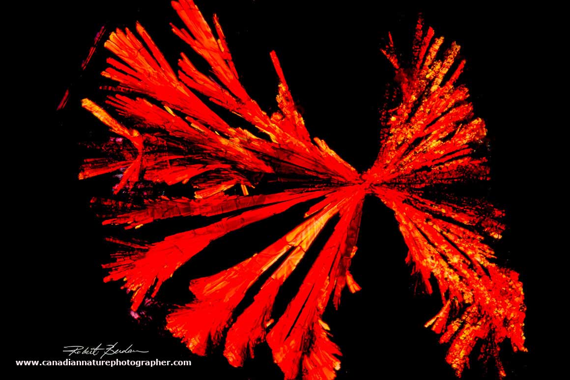
Vitamin B12 crystals that formed under the coverslip and viewed by polarizing light microscopy 400X.
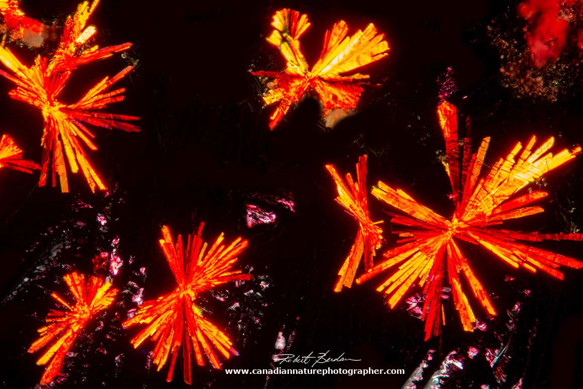
Vitamin B12 crystals formed needle-like clusters from the injectable vitamin B12 solution. Polarized light microscopy 200X.
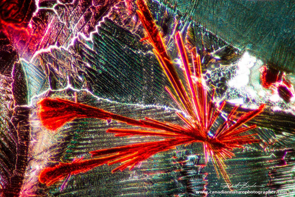
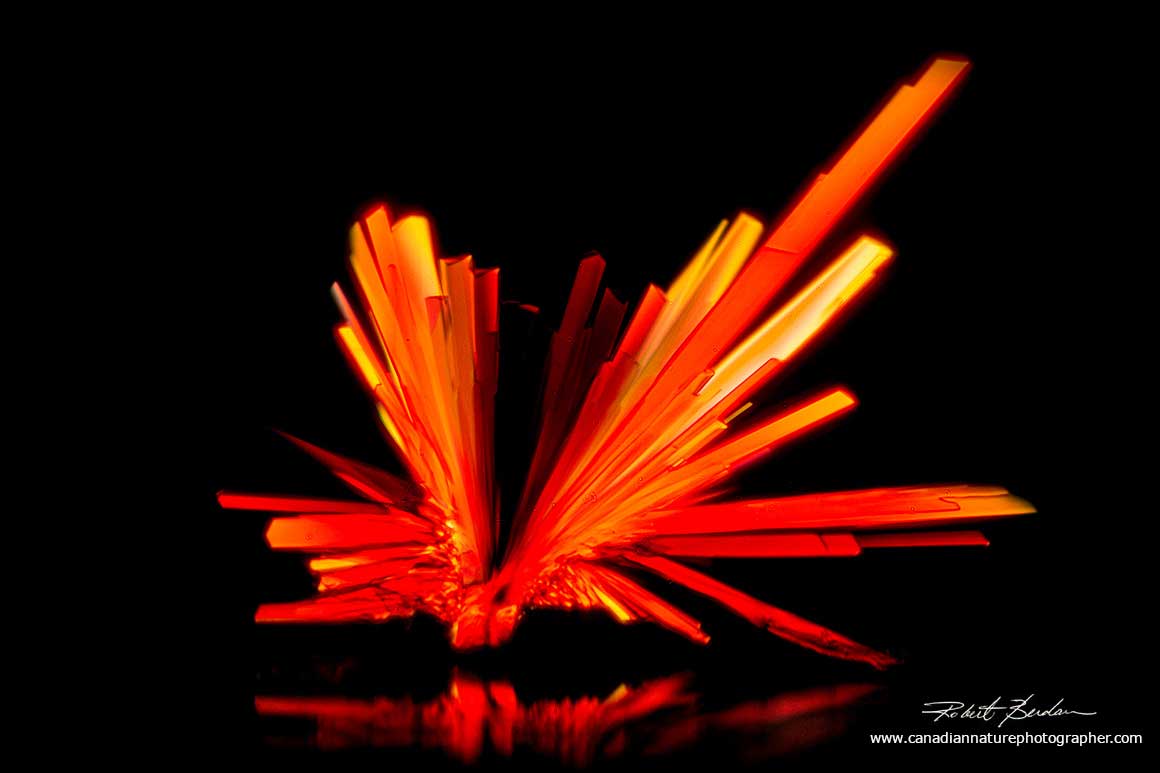
Vitamin B12 crystal that was star shaped had some long needle-like crystals. Surrounding crystals appear to be dried salt (sodium chloride). DIC microscopy 400X.
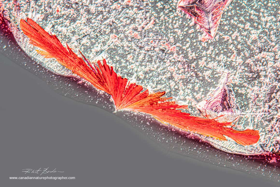
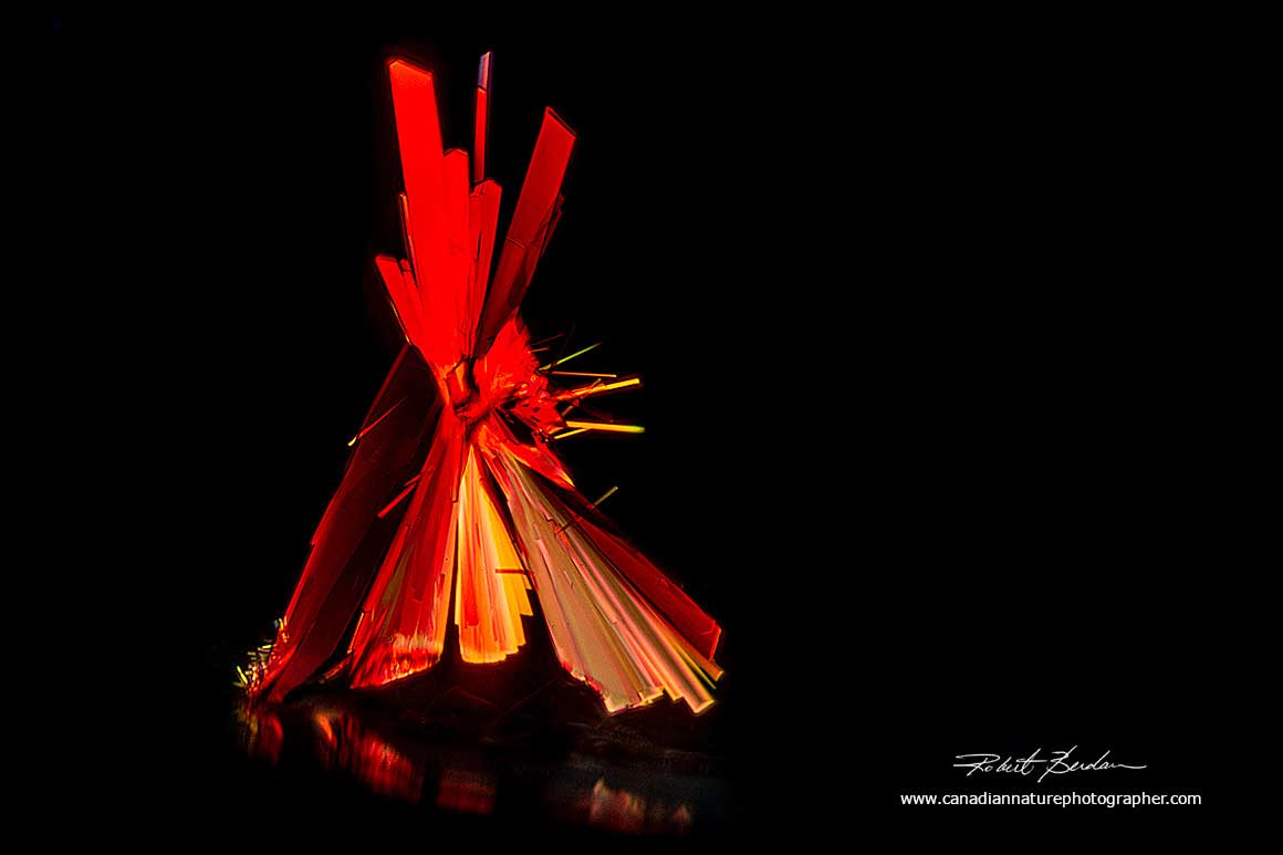
Red vitamin B12 crystal that formed on the edge of a dried drop of vitamin B12 (injectable) by DIC microscopy 200X.

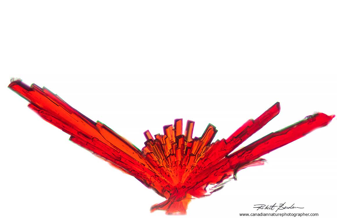
Small red flower-like crystal of vitamin B12 surrounded by salt crystals DIC microscopy 200X.
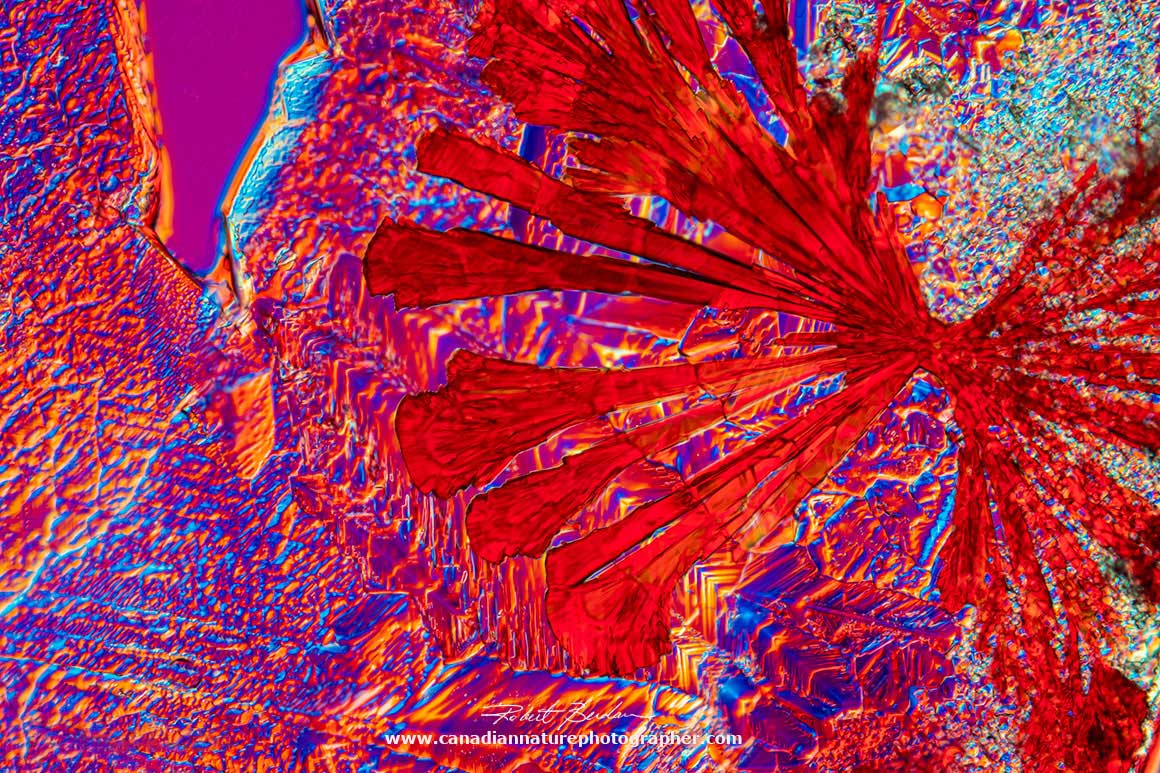
Large star-shaped red crystal of vitamin B12 by DIC microscopy 400X.
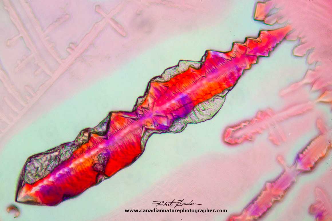
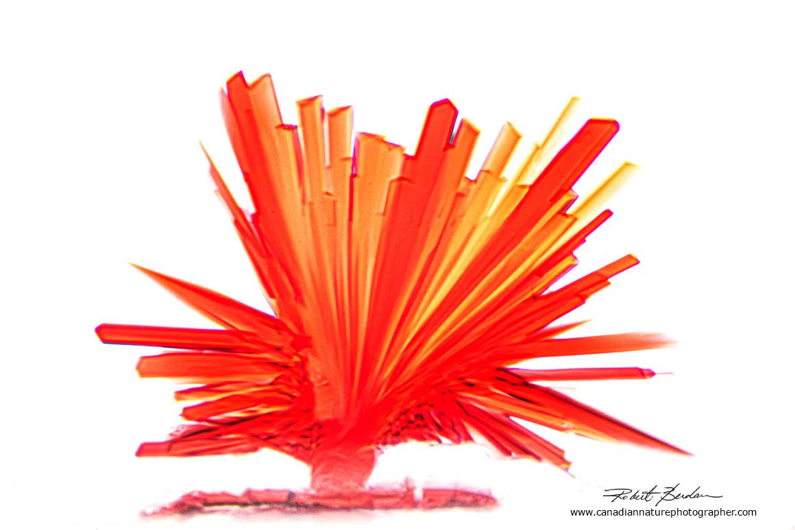
Above shows a single red crystal in a spike-shape with a thin layer of translucent material that might be sodium chloride (salt). Bright-field microscopy 400X.

Above shows a similar vitamin B12 crystal by Differential Interference Contrast (DIC) microscopy 400X.
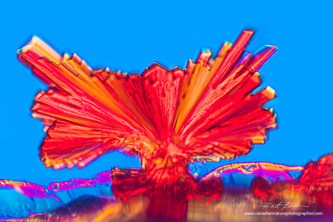
Vitamin B12 crystals on the edge of a coverslip by DIC microscopy 200X.
Salt Crystals
Sodium chloride in solution when dried on a microscopy slide can form a variety of patterns. The large salt crystals appear cubic or pyramidal in shape. Some of the crystals can form longer filaments and branches. Examination of human tear drops also exhibit similar patterns of sodium chloride when dried (R. Berdan, 2019).

Drop of vitamin B12 fluid on a microscope slide. Crystals appear similar to that found in human tear drops and are likely from salt in the saline solution. 40X Darkfield and Rheinberg lighting.

Close up of dried salt crystals (sodium chloride) from injectable vitamin B12 solution 100X.
Crystals of salt (sodium chloride) formed in a vitamin B12 solution heated and dried on a microscope slide. Differential interference microscopy 200X.

Dried salt (sodium chloride) formed in parts of the solution of injectable vitamin B12. Darkfield and Rheinberg microscopy 200X.

Salt crystals(sodium chloride) dried from a solution of vitamin B12. Darkfield microscopy 400X.
Salt crystals (sodium chloride) dried from a solution of vitamin B12. Darkfield microscopy 400X.

Salt crystals (sodium chloride) dried from a solution of vitamin B12. Darkfield microscopy 400X.

Salt crystals (sodium chloride) dried from a solution of vitamin B12. Darkfield microscopy 200X.

Sodium chloride crystals (salt) in solution of vitamin B12 with Darkfield and a red Rheinberg filter 100X.

Above image shows table salt (sodium chloride) crystals A) by bright field microscopy B) between crossed polarizers and C) salt viewed between crossed polarizers and full wave retardation plate - 50X. Salt is isotropic and transparent in polarized light even when adding a full (550 nm) retardation plate into the light path will not show additional colours as it would for birefringent crystals. Some of the bright areas in the salt crystals in B are due to refracted light from flaws in the crystals.

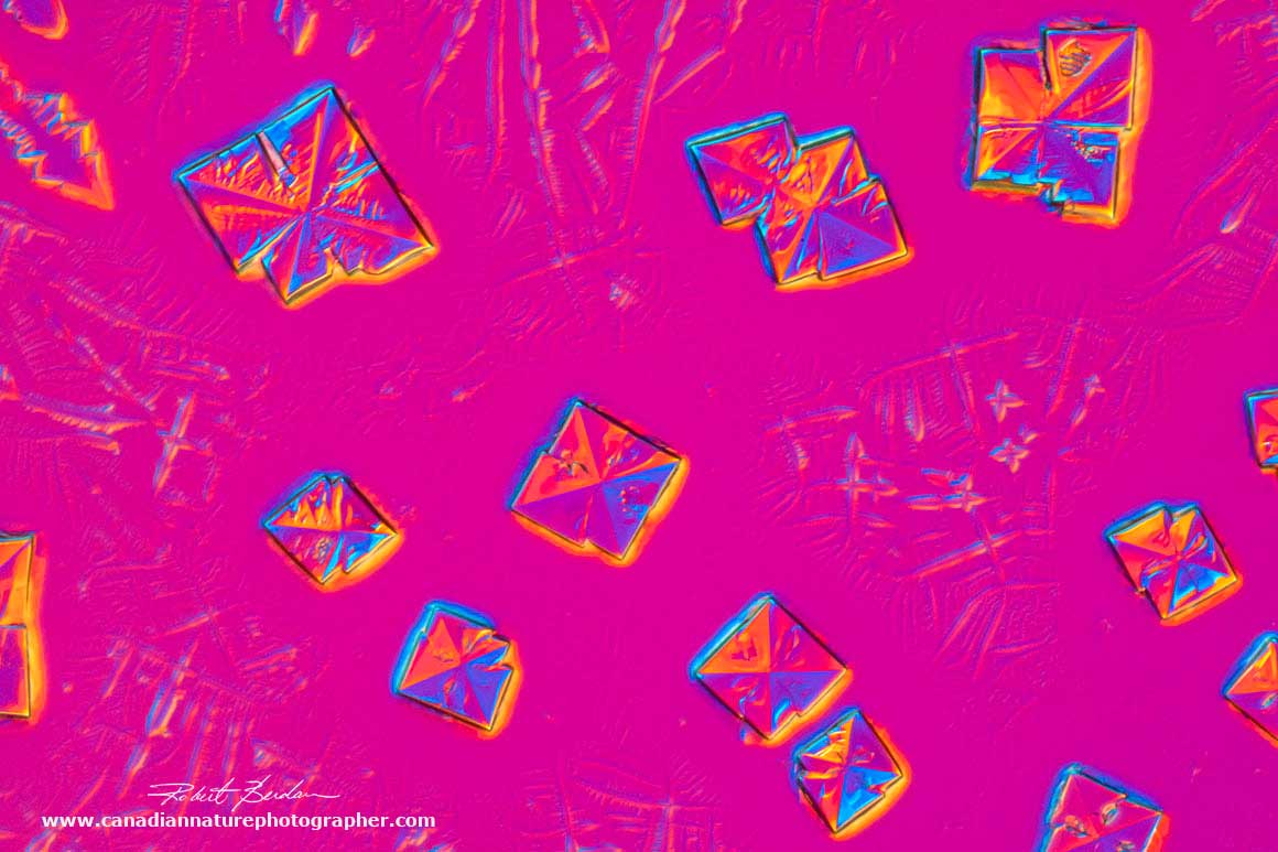
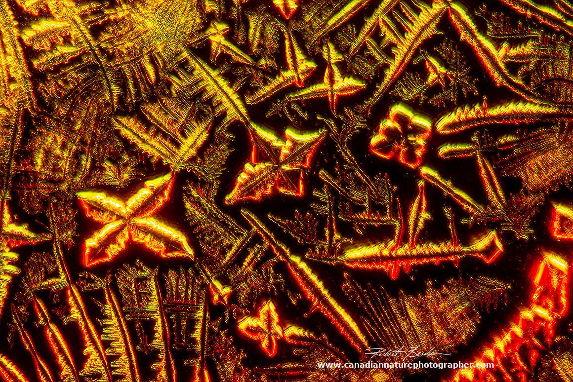
Crystals of sodium chloride by bright-field microscopy from the injectable vitamin B12 solution had red coloured components attached to them. Also note the white cross in these pictures. 400X.
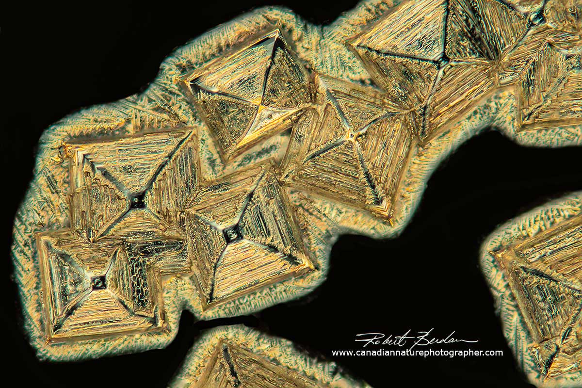
Sodium chloride crystals in water by polarized light microscopy. The crystals appear pyramidal in shape from above, but from the side they appeared cubic - 400X.
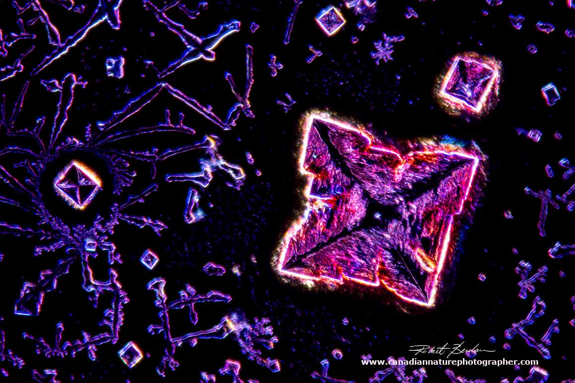
Sodium chloride crystal from vitamin B12 solution by DIC and dark-field microscopy 400X.

Sodium chloride crystals from vitamin B12 solution by DIC and Dark-field microscopy 400X.
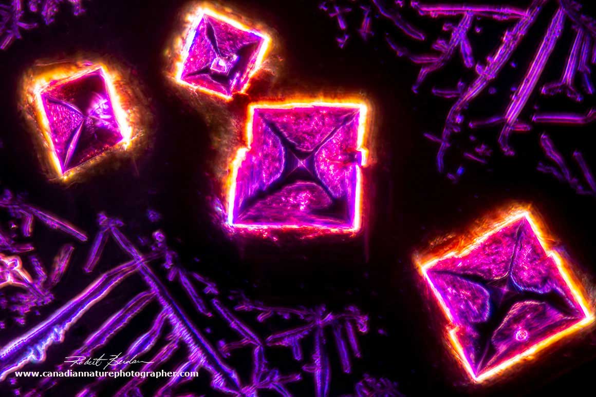
Sodium chloride (salt crystals) from vitamin B12 solution by DIC and Dark-field microscopy 400X.
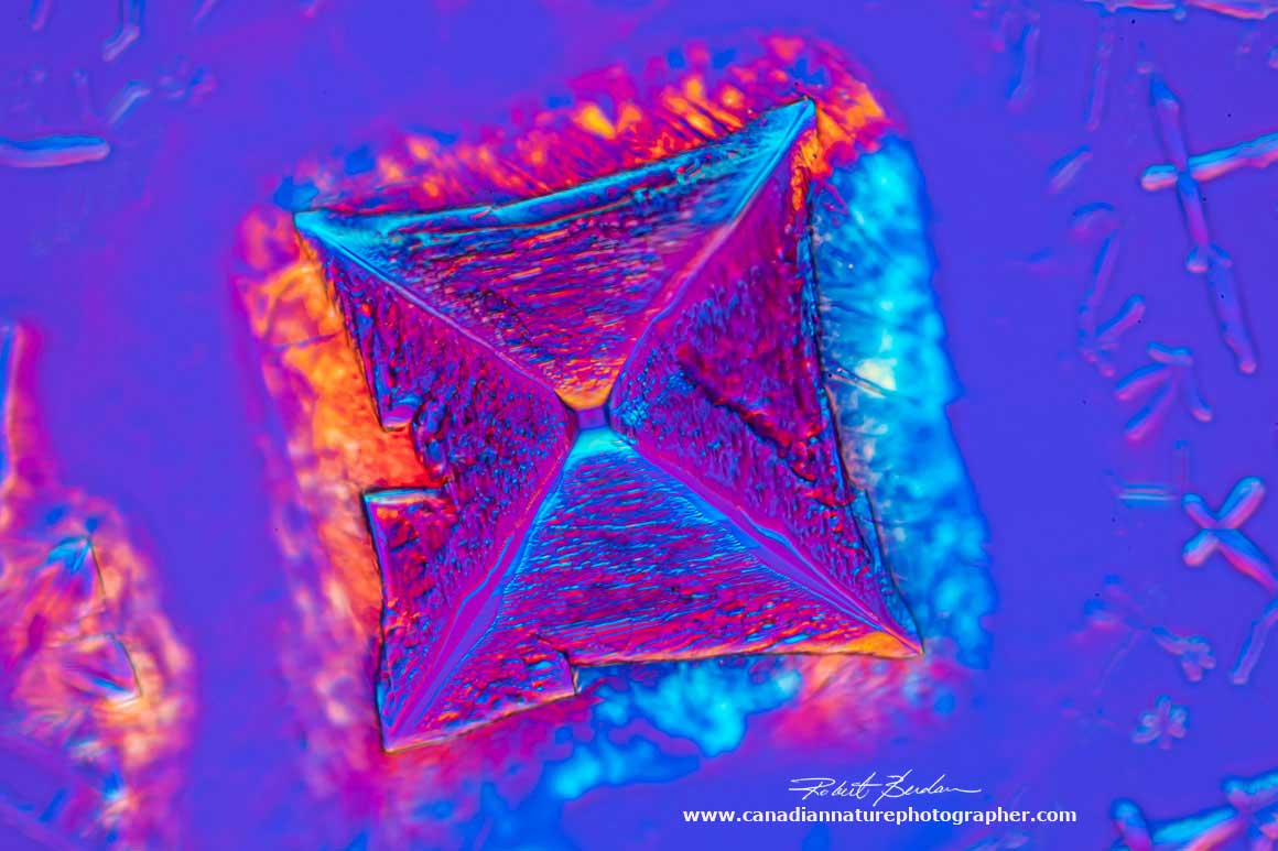
Sodium chloride crystal (salt) from vitamin B12 solution viewed by DIC microscopy 400X. In this picture the crystal appears to be pyramid shaped.

Sodium chloride crystal from a vitamin B12 solution by DIC and darkfield microscopy 630X.
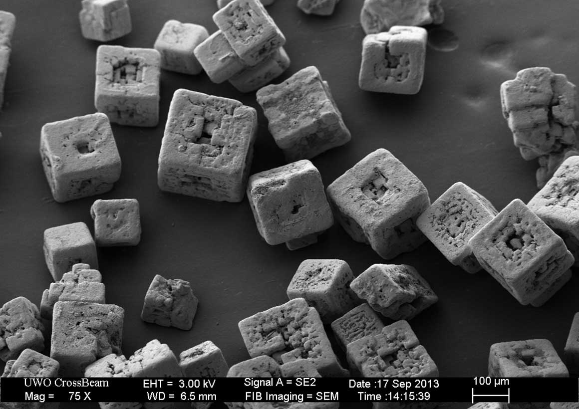
Table salt used on French Fries, scanning electron micrograph courtesy of nanofab.uwo.ca
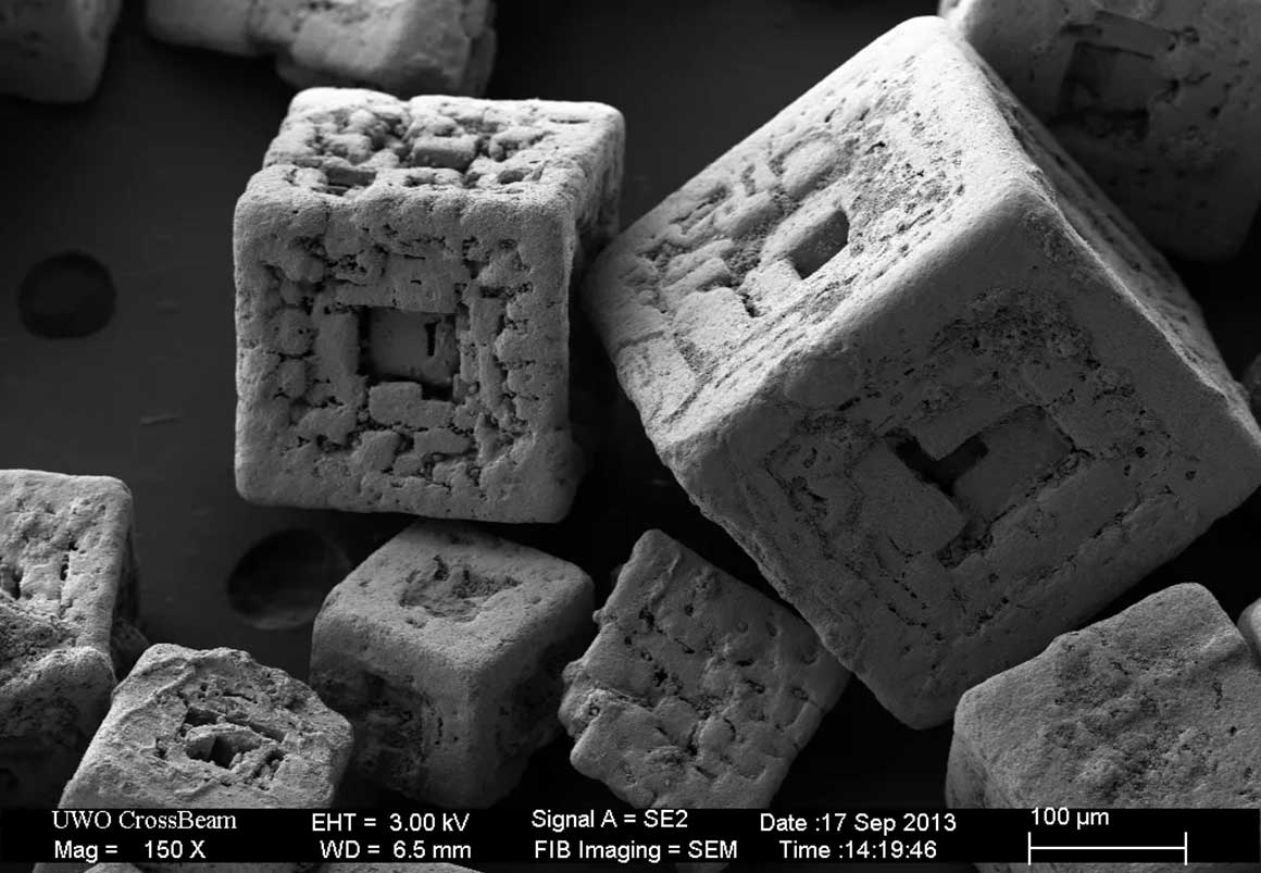
Crystals of fine salt, imaged with 150X in the FIB-SEM. Kindly provided by Todd Simpson, University of Western Ontario, Nanofabrication Facility.
Note the colours shown above in the photographs were produced by microscopy techniques and were not introduced by image processing of the images.
Discussion
Growing vitamin B12 for viewing with a light microscope from pills and also from an injectable form of vitamin B12 was not straight forward. There was lots of variability and only a small fraction of samples resulted in tiny red crystals. The best crystals grew on the inside edge of the coverslip where there was still some fluid. Growing under the coverslip allows crystals to form more slowly then those air dried.
I could find only a few vitamin B12 images on the web compared to other crystals. Some of the vitiman B12 images were not red or rod shaped as shown here. I suspect some of the photographs that claim to be vitamin B12 my be something else. I searched for a research paper showing clear photomicrographs of vitamin B12 and only found one (E. Simone et al. 2015 - readers can download PDF below in my reference section). They show vitamin B12 crystals having red needle-like crystals similar to the ones I show above.
Only the injectable form of vitamin B12 resulted in good crystals for photomicrography. Purified vitamin B12 from Sigma or other scientific supply companies might provide better results. Simone et al. 2015 report that impurities inhibited the growth of vitamin B12 which they sourced from a chemical company in China.
Other vitamins, in particular vitamin C, produce large crystals on microscope slides, some of the crystals form 2D circular shapes with a Maltese cross (R. Berdan 2019). Tylenol (Acetaminophen) forms sheets, circular and heart shaped crystals when viewed by polarized light microscopy (R. Berdan, 2017, R. Berdan 2022).
I have seen a variety of salt crystals before when added to beer (R. Berdan, 2019).Table salt photographed with a scanning electron microscope at the University of Western Ontario appear cube like and some exhibit a hole in the center. Some of appear to be pyrimidal in shape from the top. On the web I found a short article on pyramidal salt that is sold and is created through the process of solar evaporation of sea water where the water is channeled into a chain of shallow ponds or lagoons and then into large pans where the water is gradually heated, forming the pyramid shapes of the salt. The process continues until the salt reaches 3% humidity, and can take up to two years to complete - see their photos (Pyramid salt flavourfields.com). It is sometimes referred to as Cyprus pyramid salt. CanStock Photo also show pyramid shaped crystals of Himalayan salt (CanStock Photo).
Unfortuntely I did not see any side views of the salt crystals that formed from the vitamin B12 injectable solution. I need to do more experiments to see if I can find pyramid shaped crystals and photograph them from a side view. The X pattern I see on the crystals in bright field is intriguing. Lines on the salt crystals also appear to accumulate red staining material from the injectable vitamin B solution.
See "Why salt crystals grow as pyramids (sometimes) - Salt Pyramids by Adam Ragusea.The salt forms hollow pyramids called Hopper crystals. Apparently they are highly sought after by the food manufacturers. Hollow Hopper crystals taste saltier but contain less salt therefore they are healthier. Sodium chloride crystals have been grown in microgravity conditions on the space station (D. Pettit and P. Fontana (2019). These investigators produced Hopper salt cubes 2-8 mm in size. Experiments on other crystals grown in space show that protein crystals grow larger in microgravity.
I have observed that small changes in the conditions under which crystals form on microscope slides can sometimes result in large differences in the appearance of the crystals. Some crystals like vitamin C are very easy to produce. I recommend anyone wanting to make crystals for photomicrography start with Vitamin C - it is easy to work with, cheap, readily available and results in large crystals within minutes.
The objective of this study was to produce vitamin B12 crystals for photography purposes. I found that the injectable form of Vitamin B12 produced some interesting red crystals, and also some salt crystals of sodium chloride that appear pyramidal shaped from above. I need to repeat some of these experiments using pure Vitamin B12 and try to photograph the pyramid shapped crystal from a side view to see if they are in fact pyramid shaped or cubic.
Note: Educators and students may use my web images freely for reports and teaching. For commercial use please contact me. If you use my images I appreciate attribution and a link back to this web page.
Acknowledgments
I appreciate comments and proof reading by Donna, Brandon Berdan and Carl Husa.
References & Websites
R. Berdan (2019) Crystals Photographed with Polarization Microscopy: Water, Beer, Caffeine, Vitamins, Amino Acids and Human Tears
E. Simone, W. Zhang and Z. K. Nagy. (2015) Analysis of the crystallization process of a biopharmaceutical compound in the presence of impurities using process analytical technology (PAT) tools. Study was on vitamin B12 crystals and shows several photomicrographs of the red needle-like crystals. Download PDF - they show clear pictures of the crystals. Book John Wiley
Jangwoen Lee et al. (2016) The Vitamin B12 Analog Cobinamide Is an Effective Antidote for Oral Cyanide Poisoning. J. Med Toxicol 12:370-379. NIH
Fang, H., Kang, J. & Zhang, D. (2017) Microbial production of vitamin B12: a review and future perspectives. Microb Cell Fact 16, 15. https://doi.org/10.1186/s12934-017-0631-y - download PDF
Z.Schnieder (1987) Chapter 5 Purification and Estimation of Vitamin B12 in Comprehensive B12.
degruyter.com
O. Soave and C.D. Brand (1991) Coprophagy in animals: a review Cornell Vet: 81:357-364.
NIH library
Wikipedia (2022) Vitamin B12 total synthesis.
A. Mealppioni (2021) Sodium chloride (Table salt - NaCl) crystallization from solution, timelapse under polarized light - Youtube video
Microscopic images of sodium chloride crystals Wikimedia Commons
Adam Ragusea Why salt crystals grow as pyramids (sometimes) - Youtube
D. Pettit and P. Fontana (2019) Comparison of sodium chloride hopper cubes grown under microgravity and terrestrial conditions. npj Microgravity, - Open access PDF
Vitamin B12 - Wikipedia
R. Berdan (2017) Rheinberg filters used in microscopy.
Other photomicrographs of Vitamin B12 on the web
Vitamin B12 - Nikon Small world competition photos by Stefan Eberhard
Molecular Expressions - Vitamin B12
Intercell Pharma - a DIC microscope image of Vitamin B12
Dennis Kunkel - fine art america
Michael Davidson - science photo library
Sidney Moulds - Science photo library - the crystals appear similar to ours
Authors Biography & Contact Information
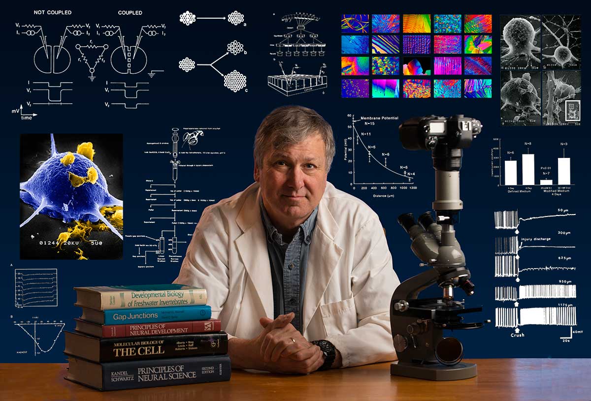
Bio: Robert Berdan is a professional nature photographer living in Calgary, AB specializing in nature, wildlife and science photography. Robert retired from Cell\Neurobiology research to pursue photography full time many years ago. Robert offers photo guiding and private instruction in all aspects of nature photography, Adobe Photoshop training, photomicrography and macro-photography. Portrait of Robert by Dr. Sharif Galal showing some examples of Robert's science research in the background.
Email at: rberdan@scienceandart.org
Web sites: www.canadiannaturephotographer.com
www.scienceandart.org
Phone: MST 9 am -7 pm (403) 247-2457.