
Microscopic Life in Ponds and Rainwater
by Dr. Robert Berdan
June 6, 2017 (Updated June 21, 2017)

Above is a rotifer, they are made up of about 1000 cells, about 0.1 mm (100 microns) in length. When a pond dries up they can form cysts which can survive without food or water for more than a decade. When wet they once again come out of hibernation. These animals have two wheels covered in hair (cilia) they use to feed and swim. 400X.

Collecting rainwater from my roof in order to examine the microscopic life that lives in my gutters
My interest in photography started with a microscope as I wanted to share images of this incredible world with others. It may look like pond scum until you look at it through a light microscope. In my previous article I showed a sampling of images taken through the microscope over many years. I got so excited about it that I began to investigate this unseen world again. The amazing thing is that there are thousands of unusual animal and plant forms and all of them can be found in your backyard, bird bath or a nearby pond or stream. In this article I show some animals that I collected near my home in Silver springs Calgary and even in rain water collected from my roof.
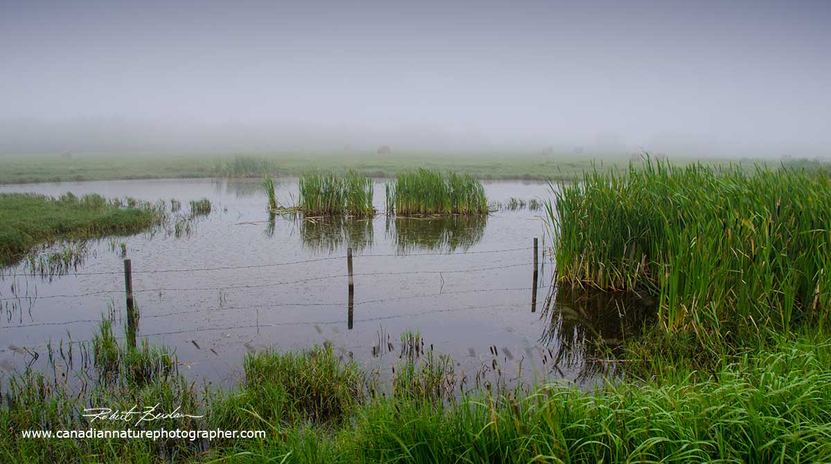
Small pond and marsh north of Calgary - an excellent place to begin searching for aquatic micro-organisms.
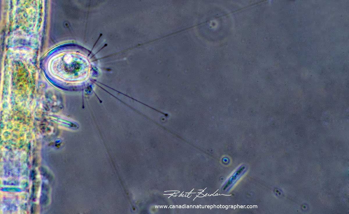
Some weird and wonderful creatures can be discovered, this protozoan (Suctorian - Podophrya sp ?) is attached to algae and has long spikes (axopodia) that it extends out in the surrounding water. 400X Phase contrast microscopy.
Euplotes is another unusual but common single celled ciliate found in pond water. 400X Phase contrast microscopy.
If you don't own a microscope, you can start examining some of the smaller plants and animals using a hand lens. If you have a macro lens you can photograph some of the bigger insects. A decent microscope can be purchased for about $100 used, check out Kijjii and E-bay - though it helps to know someone who can guide you if its your first purchase. I recently purchased another used microscope off Kijjii and with some loving care I am restoring this microscope to take pictures.
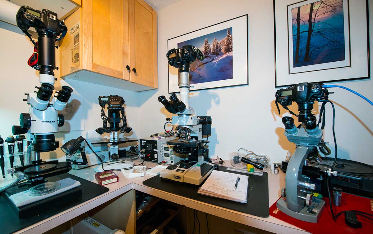
My microscopes include a Wild Stereo scope on the left capable of 5-50X, just right of it is a Canon 5D Mark II with Canon's MP-E65 mm F/2.8 macro lens with flash that allows me to take pictures from 1-5X. In the middle is my refurbished Nikon Optiphot with polarizing optics, negative phase contrast, epiflourescence illumination, and Hoffman modulation optics (allows me to examine unstained specimens). This microscope provides 4X-1500X magnification and I purchased it used from Kijjii. On the right is my first microscope - Olympus E equipped with polarization, Darkfield and positive phase contrast - it is over 40 years old and still allows me to take pictures.
It's easy to attach even a cell-phone or other camera to the microscope eyepieces. I use special photo tubes. Used and new photo-tubes can be purchased on E-bay - I purchased a new AMScope CA-NIK-SLR Camera adapter for only $88.50 on E-bay, but you can also find used ones cheaper. See this article on how to attach your smartphone to a microscope. If you can get your hands on a microscope you can take pictures. I purchased a used scope for my son for only $100. Note to see many of organisms like I show below your microscope will need to use Darkfield or Phase contrast to see many of the transparent specimens.
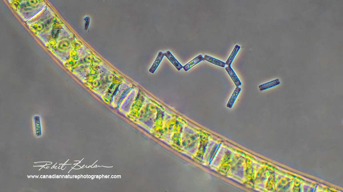
The large filament is green algae (Spirogyra) that looks like fine green horse hair when viewed in a pond - see 3rd jar from the left below to see its fine appearance macroscopically.

Some typical water samples. On the left the large jar is rainwater collected from my roof. The other samples are from ponds and streams. Anyone can collect water specimens in small jars and take them home to view.

Ciliate (Holosticha pullaster) beside algae strand, 400X Phase contrast microscopy. These animals can move very fast and capture sharp pictures of them is a challenge.
The light source needs to be very bright for photomicrography. Some of the light sources that come with microscopes are OK, but if you want to make movies or use a faster shutter speed, a bright light source is essential. I have been experimenting with LED lights - tried a 4000 lumen flash light from Amazon which works pretty well. My favourite light source thus far is an old Kodak slide projector you can see in the photo near the top of the page. A cable release reduces shake due to the shutter. If you want to see the images while you photograph - you can attach your Digital SLR with a USB cable to your laptop and use a free software program called Digicam control for PC. This software supports most of the Nikon and Canon cameras. It can be used with the camera alone, or when your camera is attached to a microscope or telescope. It's easy to use and its free!
 Digicam control software captures images on your computer while using the Live View mode of your camera. Using live mode will use up your batteries faster than normal so keep spare batteries handy.
The amoeba is a single celled animal that has the ability to alter its shape, primarily by extending and retracting pseudopod. Often studied in biology class they are common in the mud at the bottom of ponds and apparently in the eve troughs of my roof. I found more than 25 of them in rain water I collected from my roof.

Amoeba from my roof top collected from rain water. Phase contrast microscopy 800X.
Below I prepared a time lapse series of photographs taken about 5 secs apart to show how an amoeba moves over the substratum.
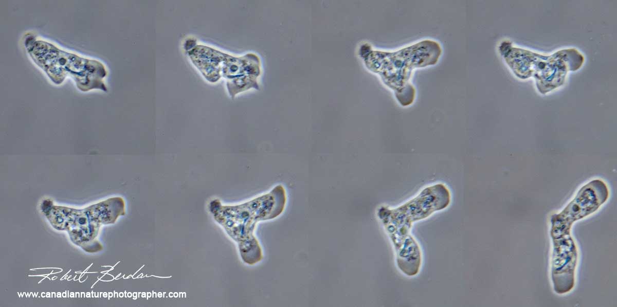
Starting at the top left and going across is a series of photos taken about 5 seconds apart showing the movement of an amoeba. I am using a technique called Phase contrast microscopy to make the amoeba visible. 200X.
Youtube video I made showing Amoeba movement - protoplasmic streaming. I increased the speed of the video by 2X and I focus above and below so you can see what is going on in all parts of this one celled protozoan. Note that inside the ameoba is a diatom that it has injested. 400X Phase contrast microscopy. There is a smaller flagellate thatseems to be 'stuck" to the ameoba. (Ooops spelling of Amoeba not Ameoba - Wikipedia article).

Copepods are small crustaceans found in fresh water about 1-2 mm in size - side view. Their large size required that I stitch 3 pictures together in the top photo. They are also called a Cyclops because they have one eye at the front. Fish and other aquatic insects feed on them. 50X.

Above a Cyclops from above - focus stack of 6 images, note the single red eye on the front of the head.

Nauplius larvae of Copepod (Cyclops) shown above. Phase contrast 200X

Oligochaete worm (Chaetogaster sp?)
Ostracods are bivalved crustaceans about 0.35 -7 mm in size and can be seen without a microscope. They have a single eye, and legs which are used to swim and bring in food. Most live near the bottom of ponds and other sources of water. 50X.
Gastrotichs are peculiar animals with caudal prongs. They are covered in cilia (small hairs) with some whiskers or spines on the front. They are made up of about 1000 cells and are common in pond water. Best way to collect them is to squeeze pond plants over a jar. They can move pretty fast. 200X Phase contrast.
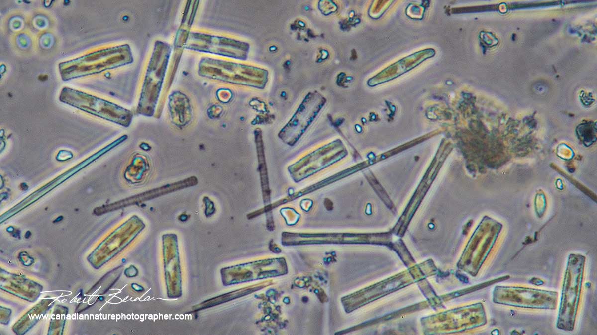


Above photos: Diatoms are small single celled plants, often in golden brown in colour. Different species are covered in delicate and intricately designed silica shells. Some species are used to test the resolution of light microscope. They move by streaming protoplasm along a raphe and seem to glide along. These photos show a number of different species. To collect them - brush the brown muck attached to rocks into a jar. Some collectors use a tooth brush. They are also common in soil and at the bottom of ponds in the mud and also in some soils. 200X.

Diatoms are the true jewels, these single-celled plants form intricate silica shells and can glide slowly over the substrate and are abundant. My best guess is this is Amphora ovalis. Phase contrast microscopy, 400X.

Single diatom with brown pigment visible. You can see some of the gratings on the silica outer shell that fits together like a soap box or pill box. This was collected from a small waterfall in Silver springs near my home. 400X
In this pond sample at the top is a filament of blue-green algae one of the most primitive organisms on the planet. Their DNA is not contained in a nucleus and they are closely related to bacteria. They can also produce potent toxins if they bloom in a pond and the toxins can kill birds and even cattle drinking the pond water. At the bottom of the photo are some diatoms - Tabularia that grows in stacks. 600X.
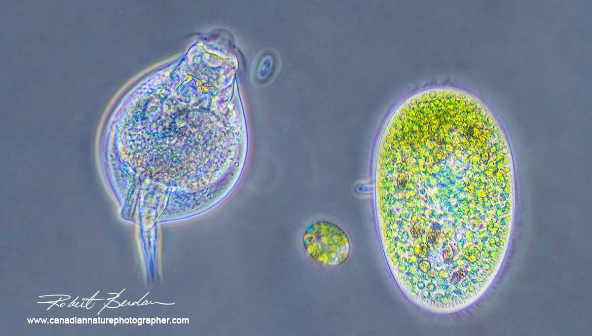
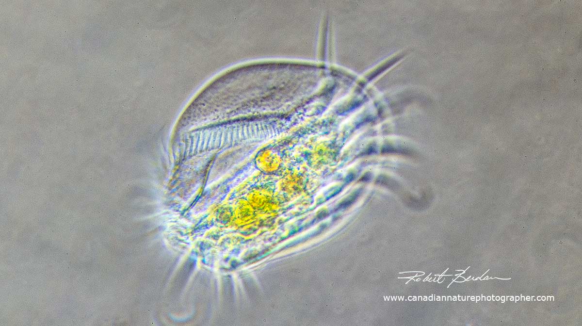
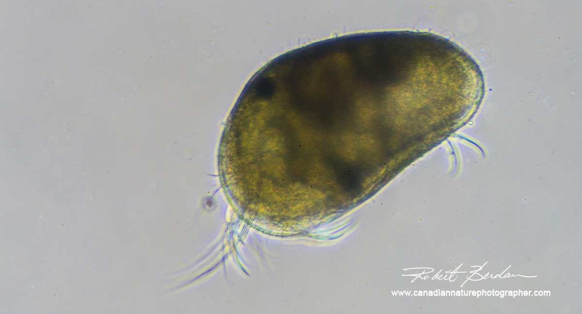
On the left is a rotifer made up of about a 1000 cells and next to it are two ciliates both are one-celled animals from pond water. The large ciliate has been feeding on smaller algae hence its green colour. Phase contrast microscopy 400X.

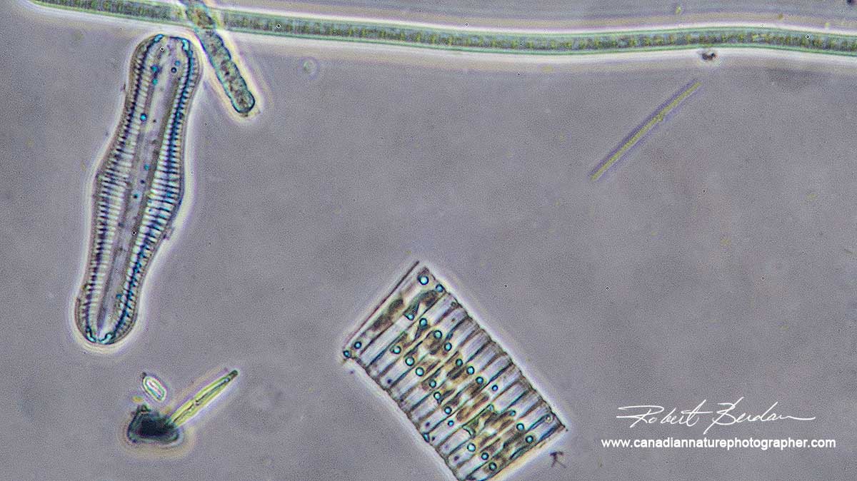
Diatoms growing on stalks attached to algae. I collected these in a drainage pool that collects storm water runoff in Silver Springs. 400X Phase contrast microscopy.

Left: Diatoms growing on a stalk attached to algae. Middle: single "S" shaped Diatom, Right: Desmid, single celled plant with bilateral symmetry. All samples are from pond water. 400X Phase contrast microscopy.

On the left is a small Diatom and the on the right is a Desmid - Closterium sp. Phase contrast microscopy 400X

Single celled ciliate with cirri (long hairs) and cilia that cover its body. These animals can move quickly. This one was collected from rain water from my roof - see top photo.

Single celled ciliates collected in rain water from my roof. They appear to contain smaller algae that they have been feeding on. 400X Phase contrast microscopy.

Stentor is a large single celled protozoan. They can get up to 0.1 to 0.5 mm (100 to 500 microns) in length. They are covered in fine hairs (cilia) that provides motility. Stentors can move about or anchor themselves to plants. This one has been feeding on smaller green algae.

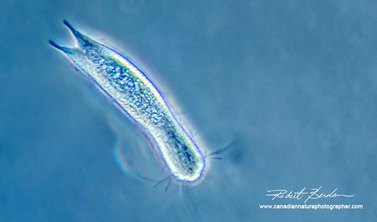
Rotifer from rain water off my roof. I had read that some rotifers live in evestroughs, but this is the first one I observed. I presume they get there by attaching themselves to the feet of birds which in turn search for food and small worms on my roof.

Small worms called Nematodes are the most abundant organisms I observed in rain water from my roof. They can be several millimetres long and are also common in soil.
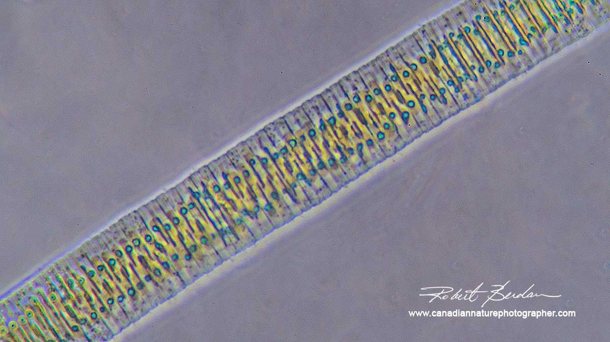
Tabularia Diatoms collected from a local pond viewed by Phase contrast microscopy. 400X
Note if you take rain water or water from a bird bath the density of organisms is relatively low, but if you concentrate them you will see many more. To concentrate the organisms you can let them settle on their own and then collect water using a pipette (can purchase them on Amazon - in fact you can purchase almost anything on Amazon!). I took 15 ml of rain water and put it into a plastic centrifuge tube and spun them for 5 minutes (I purchased a clinical centrifuge on Kijiii for $150). After removing the supernatant I examined the lower 0.5 ml which was rich in amoebas, nematodes, ciliates and other microorganisms.

Blue-green algae growing on a larger algae filament. Autoflourescence using Green Excitation. 400X Epiflourescence microscopy.

Oscillatoria is one of the most primative forms of algae, like bacteria their cells have no nuclear membrane. Some species can create potent toxins that can kill birds and cattle drinking the water. 400X Phase contrast microscopy.
Summary & Conclusion
To some it might be pond scum, but to the interested Geek these organisms are jewels. If you are interested in taking pictures with a microscope you need to do some research to find a microscope that meets your needs for an affordable price. If possible look for a microscope that has a trinocular head which allows you to attach the camera above the scope. Also there are two types of scopes 1) Stereo microscope - top photo on left. This scope is for low magnification work only (1-50X) i.e. viewing of large water bugs, minerals, flower parts etc and are they are easy to use. You can get a used stereoscope for a couple of hundred dollars 2) Used light microscopes can be purchased for about $75 and up. The more costly scopes usually have better optics and features e.g. Phase contrast microscopy. I am looking for one with Normarksi DIC (special type of contrast to view living cells) but haven't found a used one under $7,000 yet.
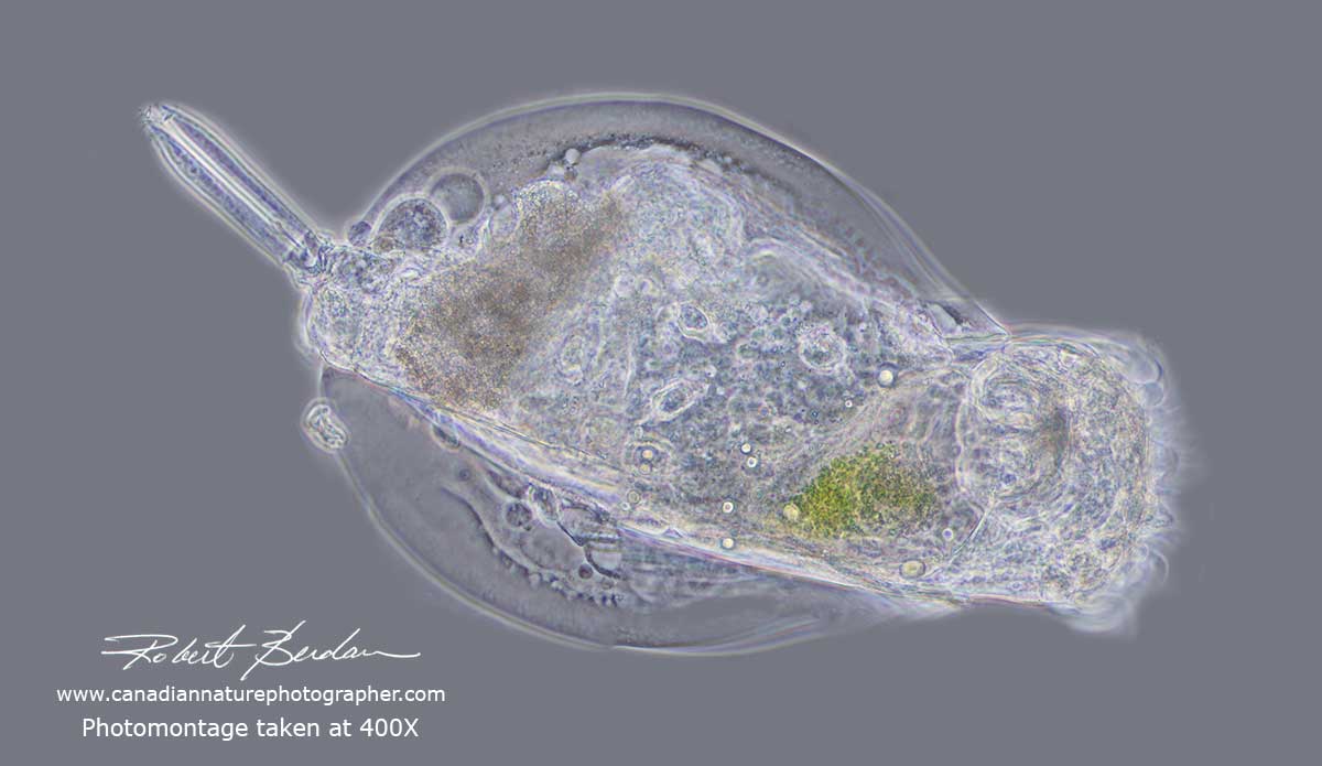
Rotifer photographed at 400X using Phase contrast, I combined 8 photos to produce this photomontage.
There are also a large number of web sites that will help you understand which microscope is right for you, but frankly you need to get some experience by working with one first (if possible) to know what to look for (check your schools, colleges etc). Below are some additional links that I hope you will find useful if you want to learn more about photo-micrography and the amazing micro-world. I will be posting more articles on microscopy in the future. If you want to get into microscopy I recommend that you start by buying a used scope. I would be happy to answer any questions you might have about microscopes and I can offer training in microscopy and photomicrography to anyone interested that lives in or near Calgary. RB
References and Additional Links.
http://greatscopes.com/microscope.htm - How to buy a microscope
http://www.olympusbioscapes.com/ - Olympus Bioscapes looking for inspiration view some of these photographs
https://westerndiatoms.colorado.edu/about/what_are_diatoms - What are Diatoms?
https://www.microscopy-uk.org.uk/mag/artjun15/sb-Diatom-Arranging.pdf - Diatom arranging Steve Beats
https://micro.magnet.fsu.edu/primer/anatomy/introduction.html - Microscopy Primer
http://www.microscopy-uk.org.uk/intro/micro.html - web site with numerous articles on microscopy
https://www.diatomsireland.com/ - Diatoms Ireland great resources web site
http://freshwaterhabitats.org.uk/habitats/pond/identifying-creatures-pond/ - Pond life
http://www.advancedaquarist.com/2002/9/breeder - growing Rotifers at home
Above are just a few of the many links on the web on microscopy
Also see my other article: Photographing Through a Microscope Photomicrography - Inner Space
Authors Biography & Contact Information

Robert Berdan is a professional nature photographer living in Calgary, AB specializing in nature, wildlife and science photography. Robert offers photo guiding and private instruction in all aspects of nature photography and Adobe Photoshop training.
Email at: rberdan@scienceandart.org
Web site: www.canadiannaturephotographer.com
Phone: MST 9am -7 pm (403) 247-2457.
Click on the buttons below and share this site with your friends