
Microscopic Pond Organisms from Silver Springs Calgary -
There is beauty in Pond Scum
Dr. Robert Berdan
May 29, 2018

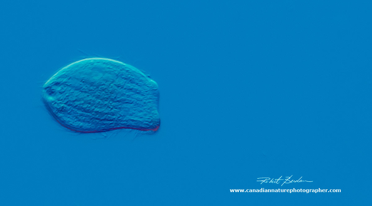
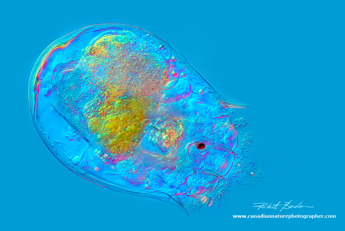
Above is a Rotifer - one of the many microorganisms collected in ponds from Silver Springs. This rotifer has a single red eye, photographed using Differential Interference Microscopy (DIC) 200X magnification.
In this article I show photographs revealing the hidden beauty of microscopic pond life found in my neighborhood ponds in Silver springs, Calgary, North West. I know that most people will never see these hidden gems unless they own and know how to use a microscope. The microscopic life forms that live in freshwater ponds, creeks, rivers and lakes is fascinating to study and these pictures show just a small sampling of the organisms. All these organisms were collected within a short walk from my house in Calgary. My assistant and wife Donna helped carry some of the sample jars and nets. One device I found very helpful for collecting specimens is a turkey baster which resembles a large eyedropper - available at Walmart for $2, otherwise a few jars, a tooth brush, a small aquarium net and some paper bags for collecting moss are all that are needed.

Storm Drainage Pond in Silver Springs, Bowmont Park, Calgary, AB
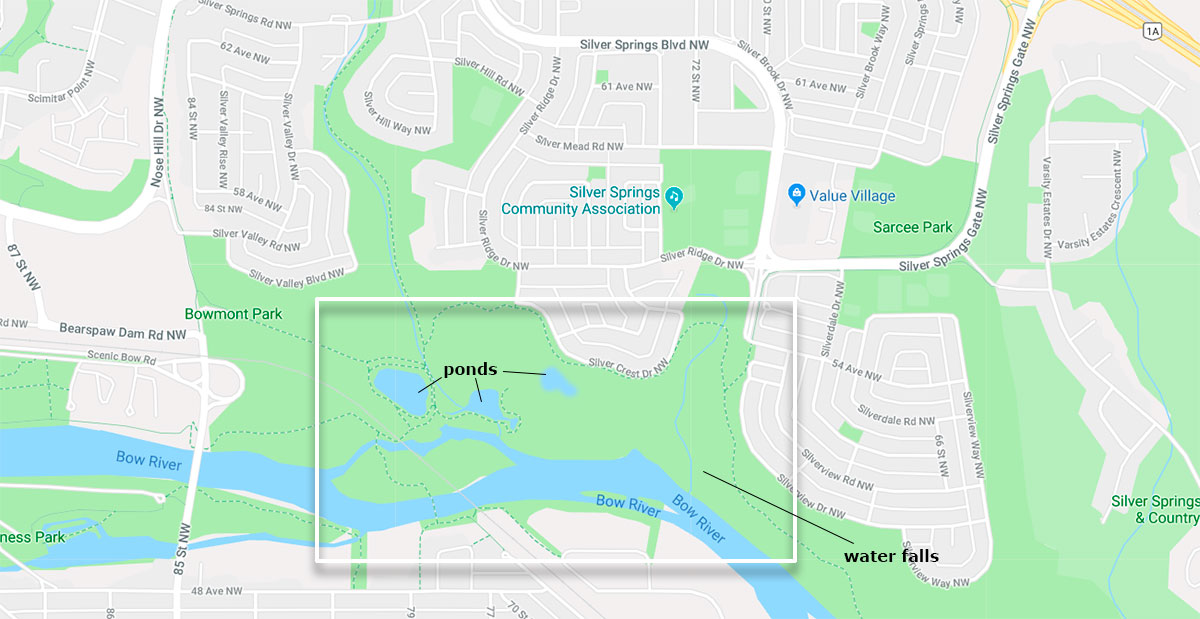
Map showing the area where the pond organisms were collected in Silver springs - Bowmont Park

Path through a coulée leading to Silver Springs Waterfalls next to the Bow river.

Just before you get to the Bow river there are small springs that form several small water falls. The waterfall is covered with brown algae and millions of Diatoms - single celled microscopic plant life that forms incredible glass (silica) shells in a wide variety of designs and patterns. Next to the falls is moss from which I collected tiny "water bears". Diatoms and other microorganisms were also be collected from under stones in the Bow river, creeks and adjacent ponds.
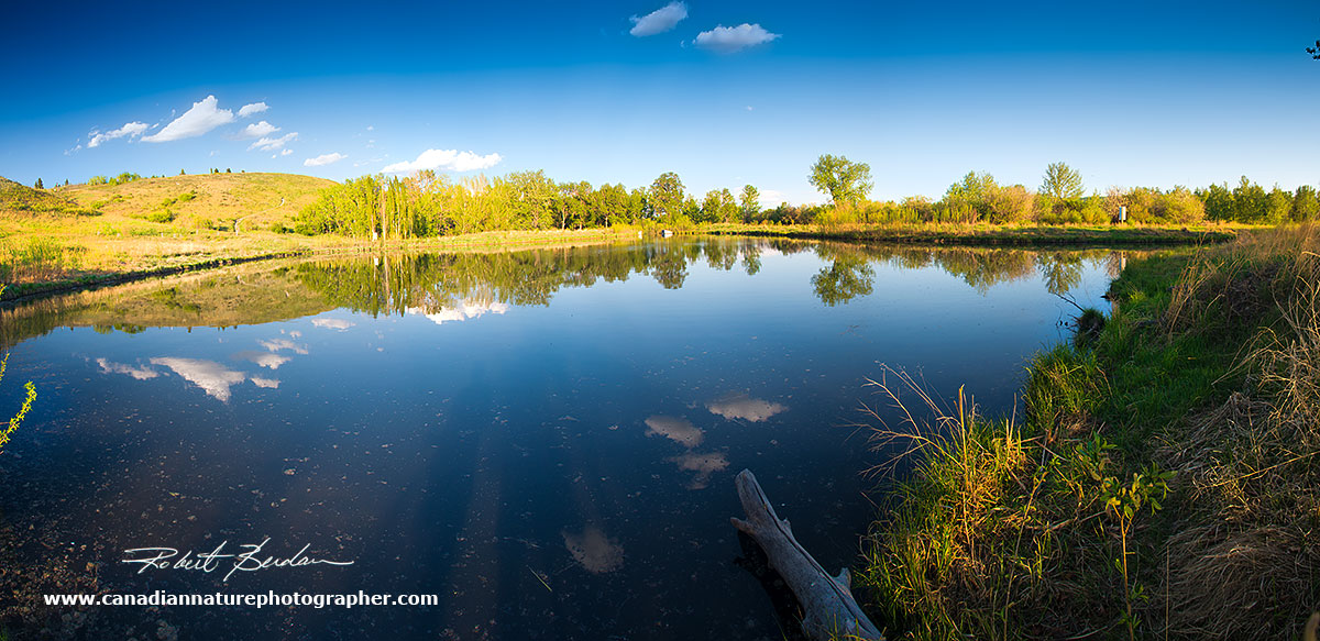
Storm Drainage Pond panoramic view - this pond usually supports ducks, blue herons, swallows and cedar waxwings as well as a large variety of algae and microorganisms shown in the pictures below.
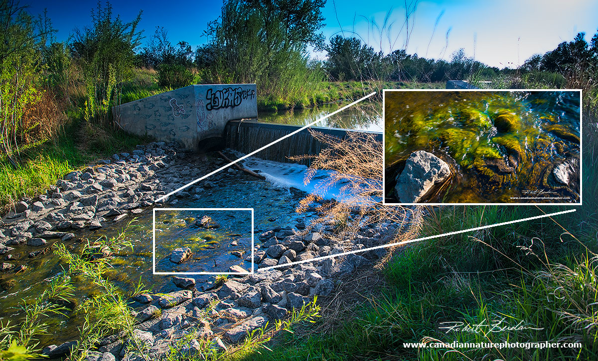
Waterfall from Storm Drainage Pond showing hair like algae composed of cladophora and spirogyra which are often associated with many microorganisms.
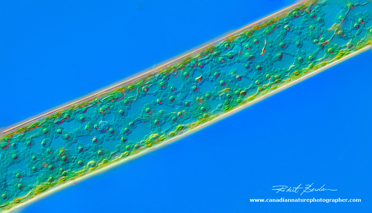
Cladophora - focus stack 200X Differential Interference (DIC) microscopy

Spirogyra algae - note the spiral chloroplast - this algae appears like fine green horse hair 200X DIC microscopy

Anabaena - Blue green algae filament. These algae are procaryotes closely related to bacteria and are some of the most ancient animals on the planet going back about 3 billion years. 600X DIC microscopy. Some of them can form potent toxins which can kill birds and livestock.
Blue-green algae - Oscillatoria 600X DIC microscopy
Protococcus algae - 600X DIC microscopy
Tribonema - unbranched filamentous algae with pale-yellow-green chloroplasts. The filaments fragment resulting in H-shaped sections. Tribonema is one of the first of the algae to appear in ditches and swamps after ice thaw.
Diatoms
Diatoms are single celled plants that form the basis of the food chain and produce about 20% of earth's oxygen. These plants are encased within silica shells and can be either pennate (rod shaped) or disc shaped. There are thought to be about 2 million species. They are found in fresh water, salt water and brackish water. They are unicellular, but can form colonies. They generally range in size from 2-300 microns (one micron = 1\1000 of a millimeter). Some can move by the extrusion of protoplasm. They appear like jewels and have a life span of about 6 days. Diatoms are used in silver polish, toothpaste, insecticides and have many other uses. They require a microscope to see them.
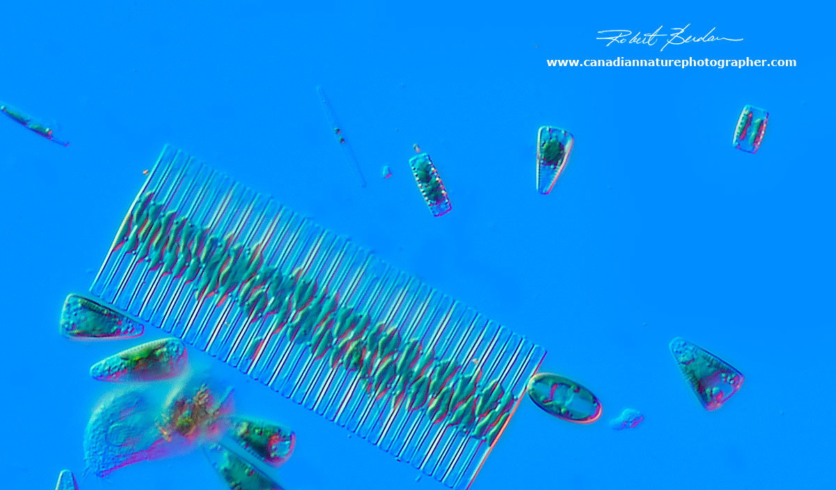
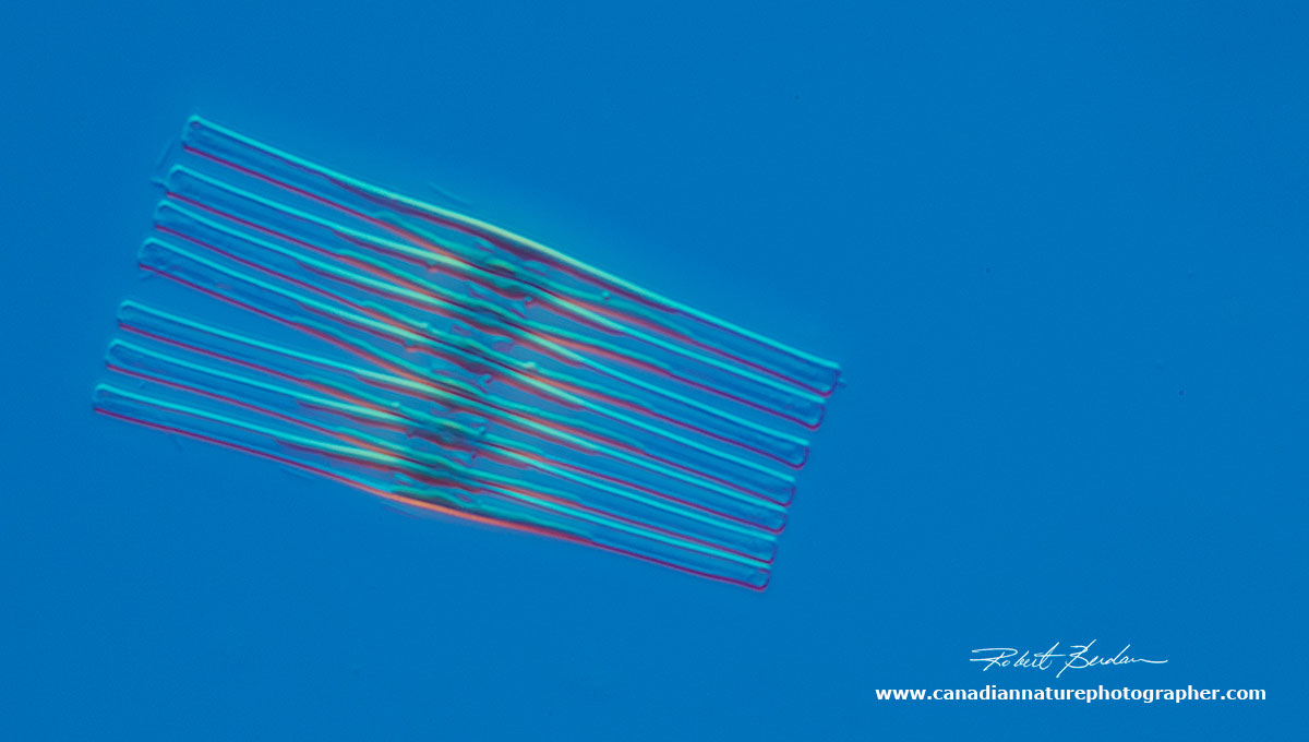
A variety of Diatoms collected at the Silver springs waterfall. The large ribbon shaped structure is a colony of Fragilaria diatoms, there are several other species also visible. 400X DIC microscopy.
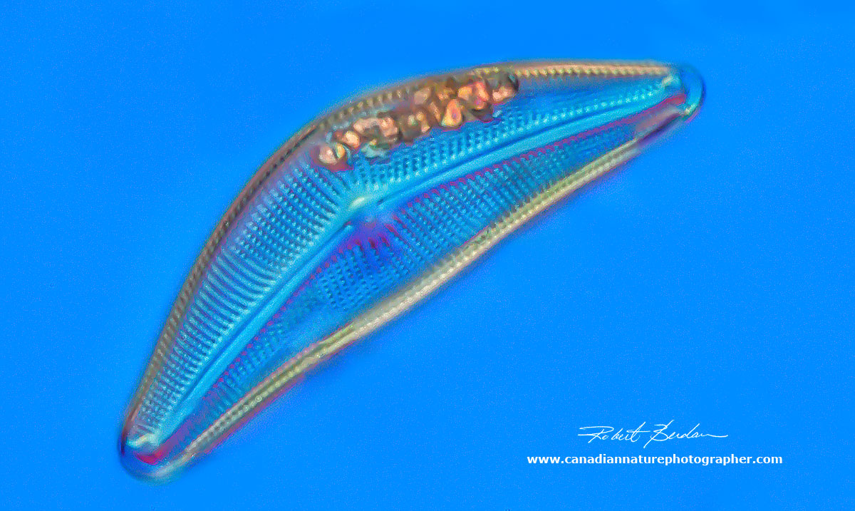
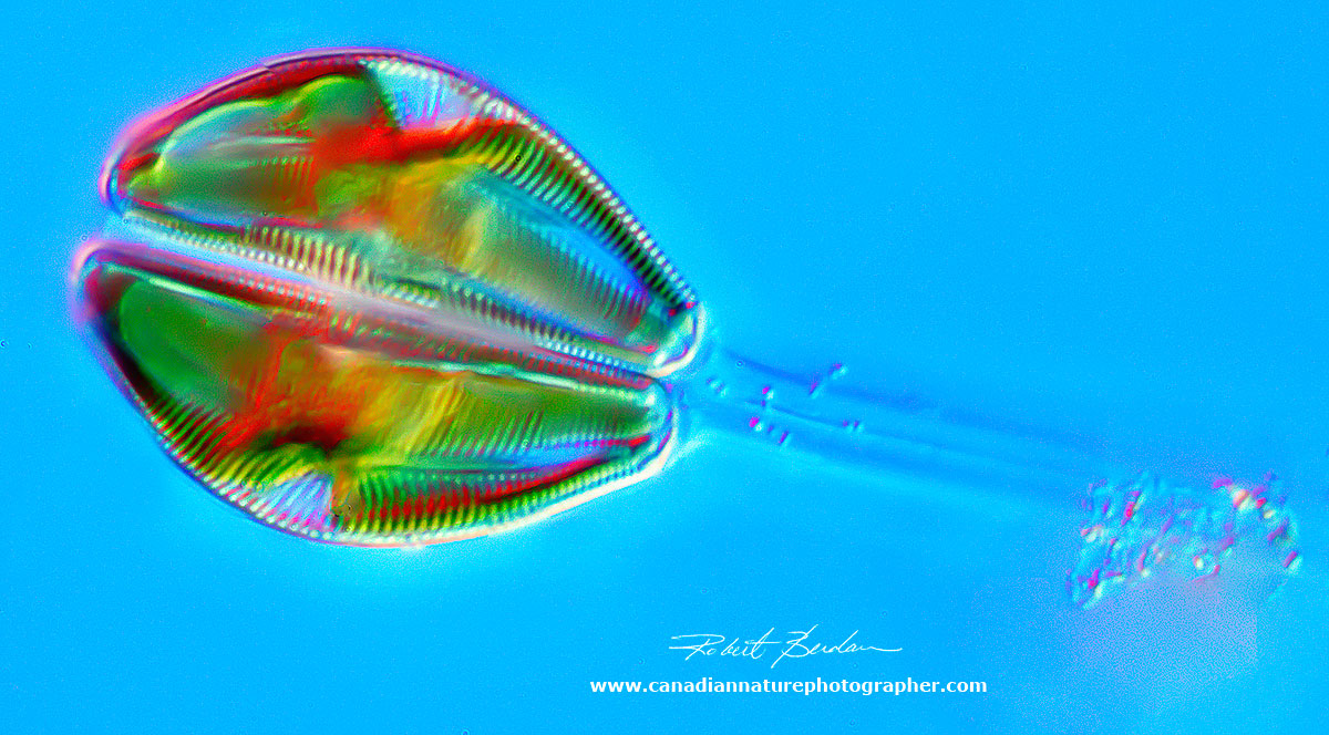
Diatom Cymbella sp. collected from the water fall. This is the silica shell only about 400X DIC

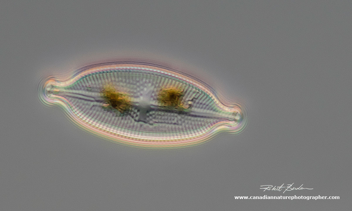
Diatom Navicula sp. note the oil droplets and brown pigment inside - Valve view (top view) 400X

Pinnularia sp. of Diatom showing oil droplets and brown chloroplasts 400X DIC microscopy.

Diatom showing a green chloroplast and oil by which it stores its food. 400X DIC microscopy.
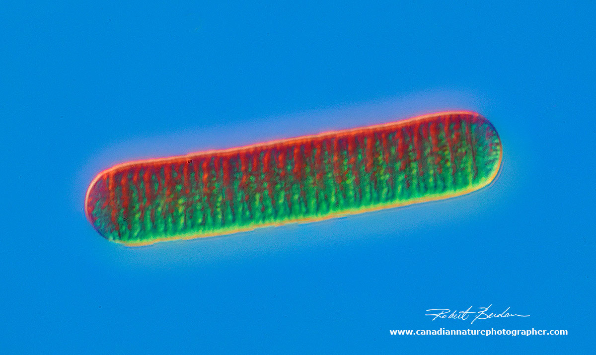
Diatom Stauroneis phoencentron Valve View 400X DIC microscopy
Nitzschia sp of Diatom 400X DIC microscopy
Nitzschia sp of Diatom 400X DIC microscopy

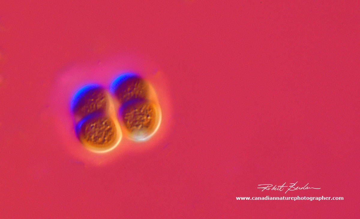
Nitzschia sp Diatom - showing two of them side by side 400X DIC microscopy

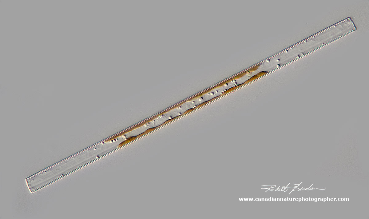
Tabellaria sp of Diatom 400X DIC microscopy

Navicula sp of Diatom showing the chloroplast inside 400X DIC microscopy

Navicula sp of Diatom - silica shell broken open and you can see the lobed chloroplast and nucleus in the center 400X DIC microscopy

Nitzschia sp of Diatom 400X DIC microscopy

Nitzschia sp of Diatom 400X DIC microscopy

Note the brown pigment in side this Diatom 400X DIC microscopy
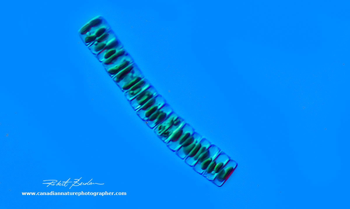
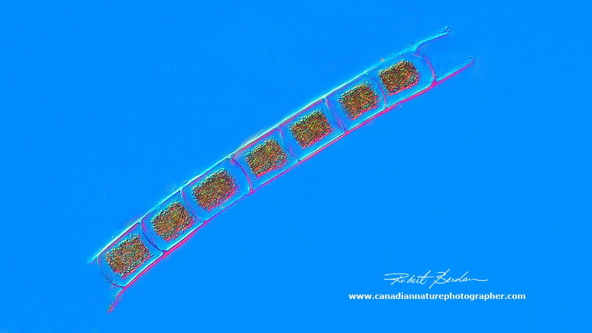
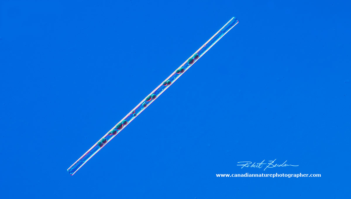
Fragaliaria sp of Diatom forming a long chain 400X DIC microscopy
Cymbella sp of Diatom growing on a stalk 400X DIC microscopy
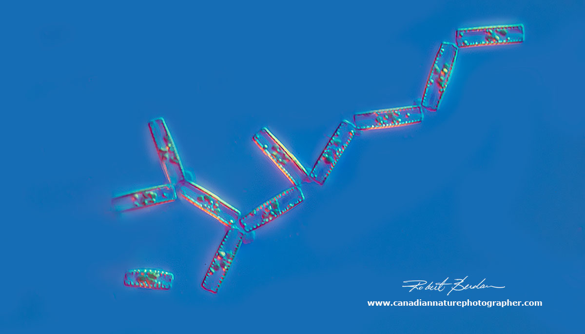
Tabellaria sp of Diatom forming a zig-zag chain - girdle view 400X DIC microscopy

Navicula sp of Diatom 400X DIC microscopy

Meridion circulare Diatom valve view 400X DIC microscopy
Diatoms Fragilaria sp. 400X DIC microscopy.
Protozoa
Protozoa are single-celled eukaryotes that feed on organic material, bacteria and other protozoa and diatoms. Protozoa are now considered to be in their own Kingdom. There are four main types 1) Sarcodina - amoeba move by streaming their protoplasm 2) Ciliates - move using cilia 3) Mastigophora - flagellates and 4) Parasitic forms - sporozoa.

Amoeba sp 400X DIC microscopy. Amoebas are found by sampling the bottom sediment.

Amoeba ejecting a digested diatom 400X DIC microscopy
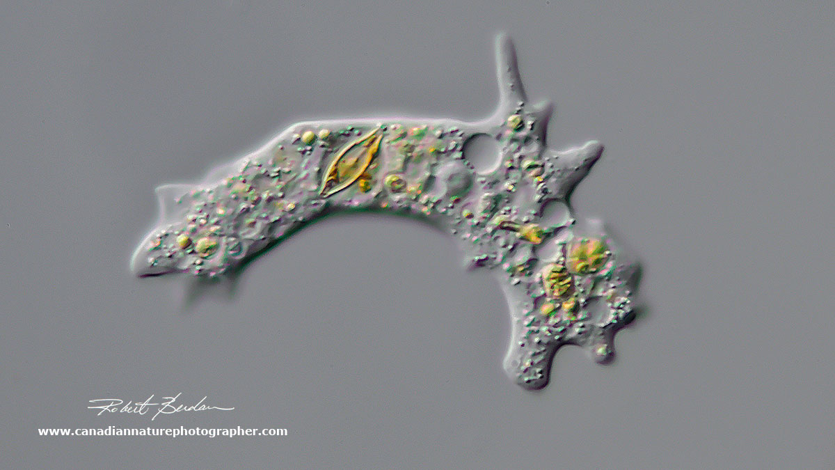
Amoeba with a diatom visible inside 400X DIC microscopy

Amoeba 400X DIC microscopy

Stentor roeselii- a single celled Ciliate that can grow up to 2-3 mm in size 100X DIC microscopy.
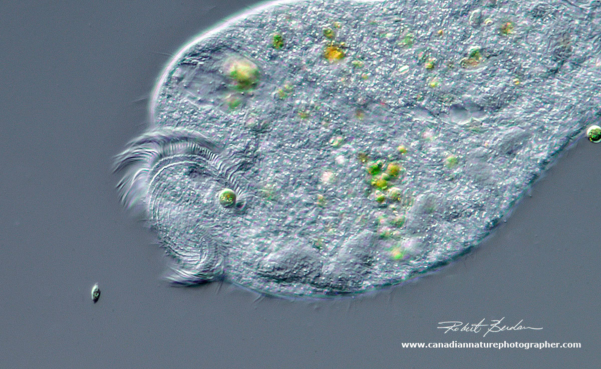
Stentor roeselii - close up of the apical end showing the cilia that brings food into its cytosome (mouth). This species of Stentor has single celled algae living inside them in a symbiotic relationship - i.e. acting like solar cells and they share the food produced. 400X DIC microscopy.
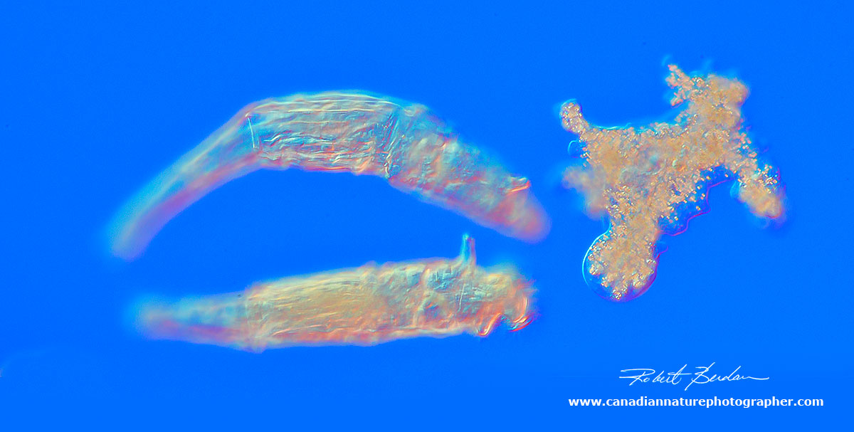
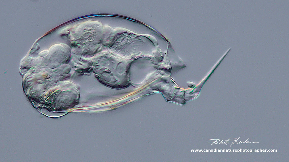
Left is a pair of Bdelloid rotifers encountering an Amoeba 200X DIC microscopy.
Ciliate - pink in colour - Blepharisma steini 400X DIC microscopy
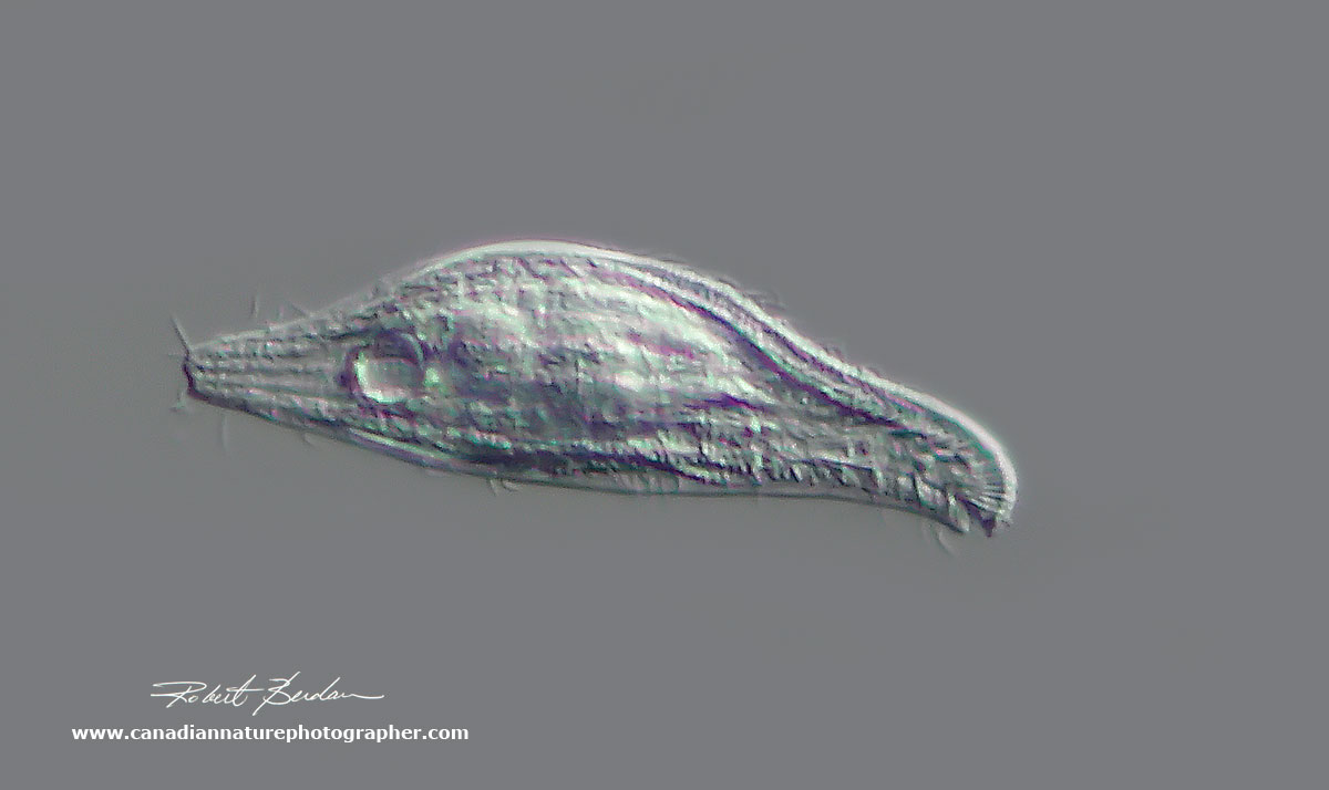
Lionotus sp? - Ciliate 400X DIC microscopy

Heterotrich ciliates possess a zone of membranelles composed of fused cilia and appear thicker - Oxytricha sp (?) 300X DIC microscopy
Oxytricha sp (?) 400X - ellipsoid body 400X DIC microscopy
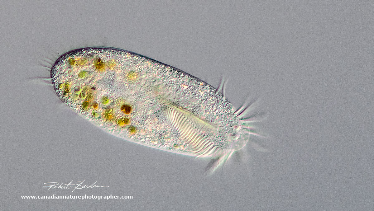
Unknown ciliate that appears to have 2 nuclei and may be dividing - 400X DIC microscopy

Unknown ciliate with green algae (Chlorella sp.) inside - 400X DIC microscopy

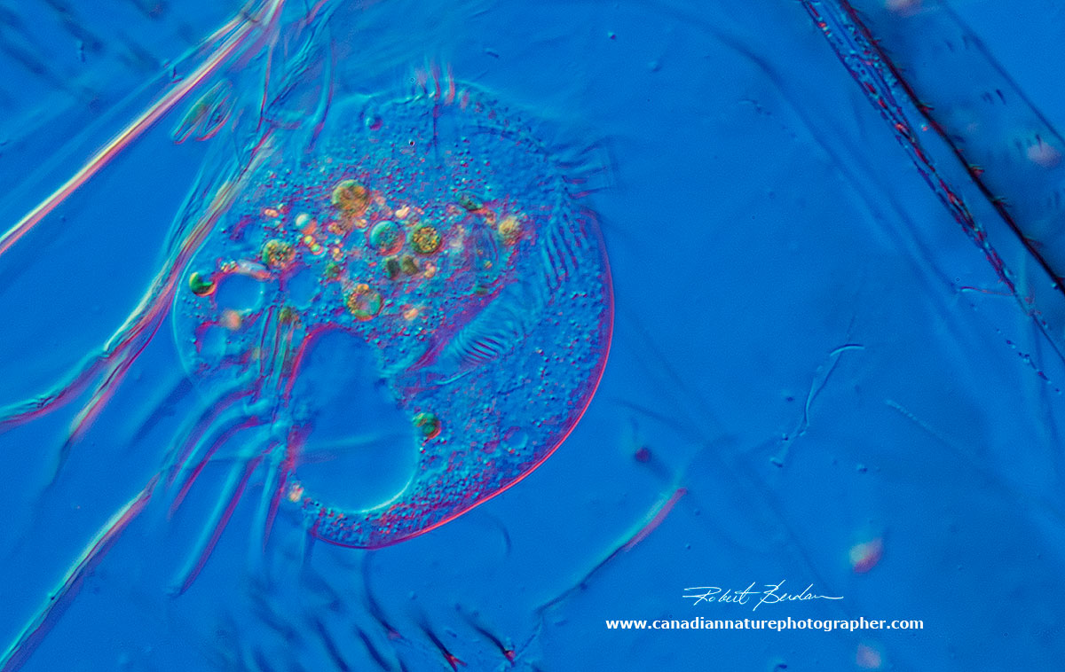
Unknown Ciliate 400X DIC microscopy
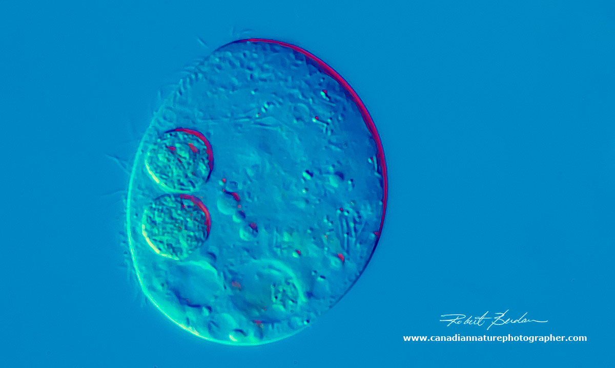
Ciliate - Euplotes sp? 100 microns in diameter - 400X DIC microscopy, this ciliate was eating inside the integument of a dead freshwater arthropod
Spathidium sp found in moss - 600X DIC microscopy
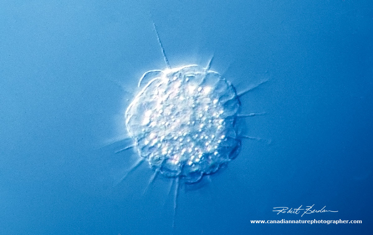
Heliozoan - sun animalcule. Actinosphaerium sp. They have radiating siliceous spines that capture their prey 400X DIC microscopy focus stack.

Flagellate - with 2 unequal length flagella, Anisomea sp (?) with one flagellum trailing - 400X DIC microscopy
Other Multicellular Microorganisms

Simocephalus sp (identified by Dr. Robert Walsh) - small crustacean about 1 mm in size. This one has several eggs visible inside. The are often called "water fleas" and are visible to the naked eye. Darkfield microscopy 50X

A closeup view of Simocephalus sp head showing it's large eye - focus stack. 150X Darkfield microscopy.

Copepod nauplius larvae, note the single red eye - Darkfield Microscopy 100X
Tardigrades - "Water Bears"
Tardigrades also called water bears are found in moss, lichens and leaf litter. I collected the moss next to the waterfall in Silver springs and I placed the moss in a dish of distilled water for several days and then collected the water and concentrated the particulate material with a centrifuge. These animals are unusual in that they can withstand dessication, high and low temperatures, and even ionizing radiation when they form a tun stage. Tuns or cysts expel most of the water from their bodies and they use a sugar Trehalose to replace the water. In this state they can survive for years and do not age as they are in suspended animation. Tardigrades have 8 appendages with claws that are important taxonomically. The water bears I photographed had 2 dark eyes on the anterior end and belong to the group of Eutardigrada. Water bears move constantly and are not easy to photograph for this reason. This one was 0.4 mm long (400 microns) and it could be seen by eye as a small speck. I used a stereoscope to find them more easily in a Petri dish filled with water whereby I then placed the specimen on a microscope slide for photomicrography.

Water bear via DIC microscopy 100X - the head is at the top left.

Water bear via bright field microscopy - 100X note the 2 eyes on the head, bottom right.

Water bear Darkfield microscopy about 200X note the eye spot on the head. You can see 4 legs on its side it has 8 in total with small claws on each end.

Dorsal view of a Water bear (Tardigrade) about 200X DIC microscopy - note the 2 eyes on the head (left side). This specimen likely belongs to the group of Eutardigrada.

Head region of a Tardigrade showing some of the cells inside called coelomocytes believed to have a defense role and the pharyngeal apparatus in the center which is important taxonomically. I have labeled the parts of the pharyngeal apparatus. The Stylets are everted during feeding and used to pierce plants. Focus stack of 8 images 600X DIC microscopy.
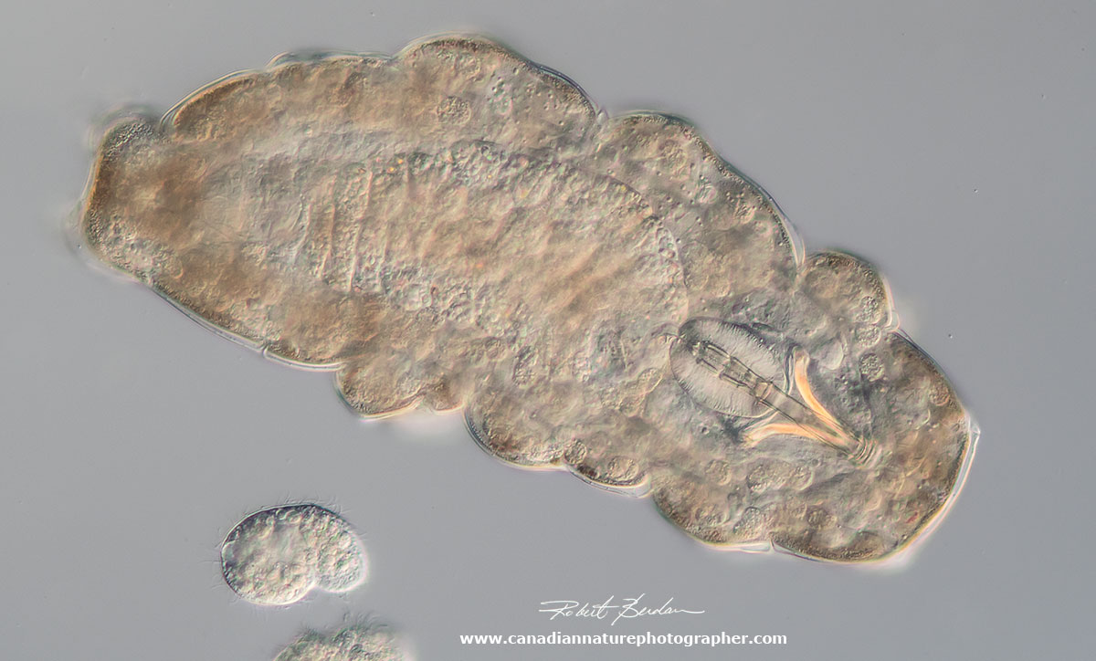
On the lower left below the Tardigrade is a small ciliate 200X DIC microscopy.
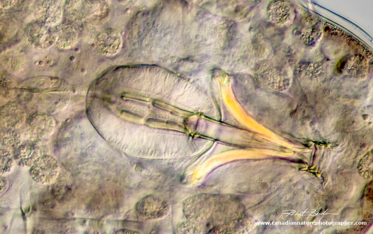
High power view 600X of the Pharyngeal apparatus near the head of the Tardigrade. DIC microscopy.
Rotifers
Rotifers are small animals sometime called "wheel animalcules" because they often have cilia on the anterior ends that appear to rotate. The cilia bring in food and can be use to propel them through the water. They are small in size about 0.1 mm (100 microns) and about 120 genera. They are well adapted to their environment and as many as 5000\litre have on occasion been counted. There a wide variety of species though identification to the species level requires considerable familiarity with these tiny animals. These animals can also form cysts when a pond dries up and survive in suspended animation for years. Some species have one or more eyes. You can see some video of rotifers in my article "Photomicrography and Video of Protozoa, Volvox and Rotifers".
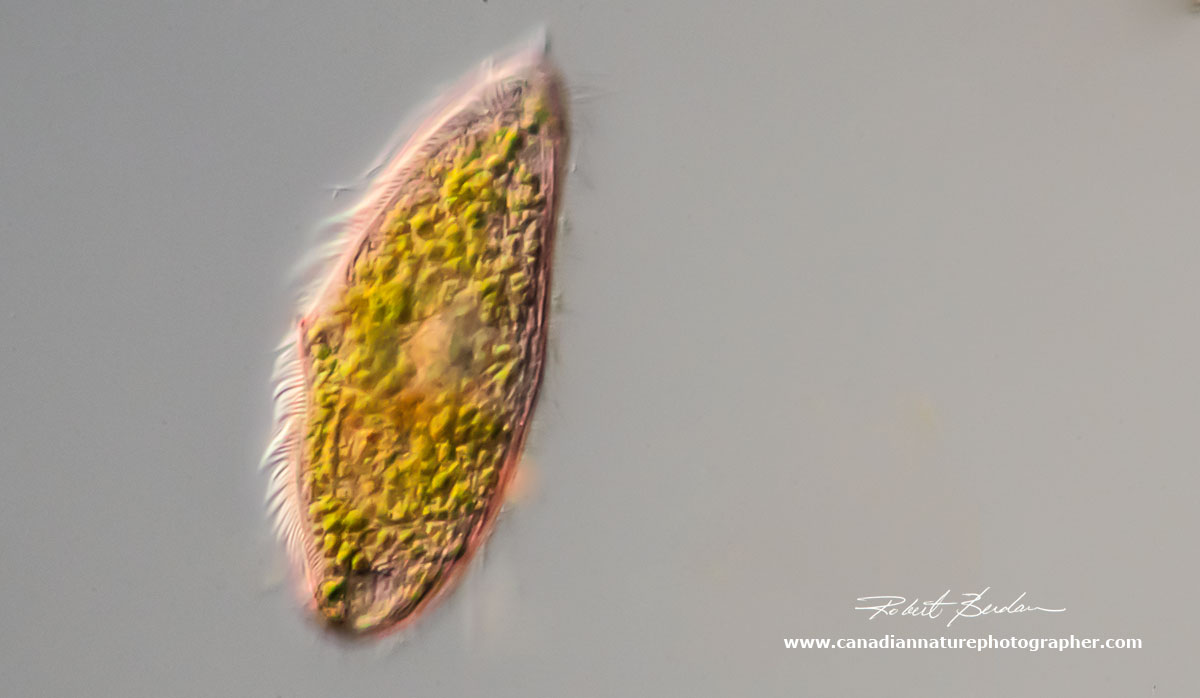
Rotifer - Notholca sp? Note the single red eye. 400X DIC microscopy
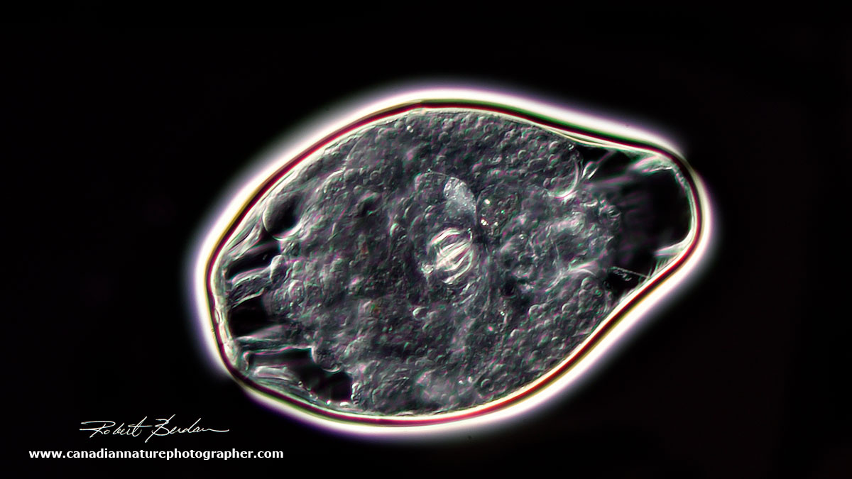

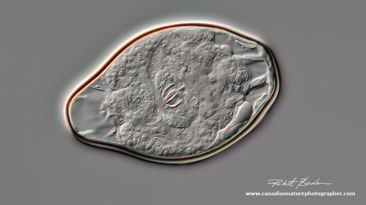
Above are three photos of a rotifer cyst whereby they can survive dessication - Top Darkfield microscopy, the bottom two are DIC microscopy, 400X.
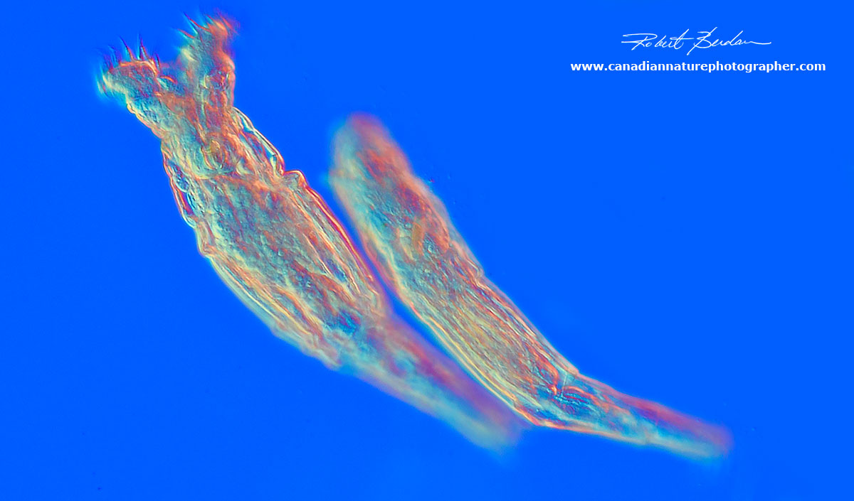
Female bdelloid rotifers. These animals consist of about 1000 cells in a syncytium. The have cilia that appear to rotate around the head that bring bacteria and other food into their digestive tract. They are about 0.1mm in length (i.e. 100 microns, one micro - 1\1000 of a millimeter). 200X DIC microscopy.
Rare male rotifer - male rotifers are rare because many females reproduce parthenogenically (without sex), but can sometimes reproduce sexually when males occur. Males often do not feed, they have only one purpose - to fertilize eggs. Males often lack mouth parts and a digestive tract and this one also had what looked like sperm inside the top right chamber. 200X DIC microscopy.
Desmids are symmetrical unicellar Plants
Desmids resemble Diatoms, but don't have silica cell walls instead their walls are made of cellulose and they form a division of the green algae. They are about 5,000 species and they vary in shape and size and are found mostly in freshwater. Desmids often take on symmetrical shapes and the largest ones can just be visible to the naked eye. Some species are motile.
Desmid Pleurotanium sp.- focus stack 200X DIC microscopy.

Desmid Closterium sp focus stack 200X DIC microscopy. This species is motile and can pivot about due the extrusion of mucilage from pores in the outer cell wall. Note the nucleus in the central region.
Worms
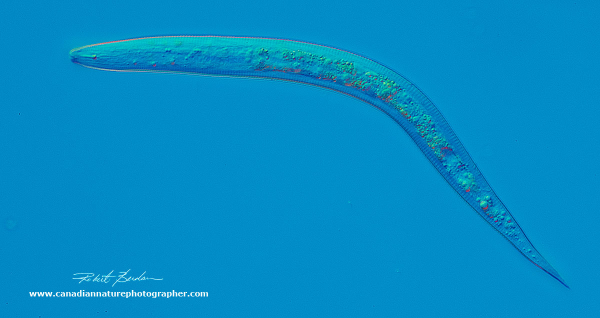
Nematodes are small transparent worms common in soil and freshwater 200X DIC
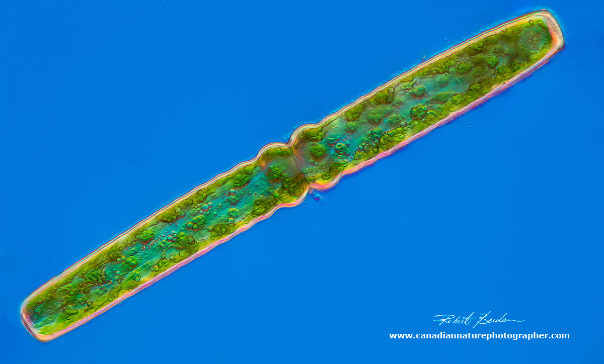
A bristle worm, Nais 20X DIC Microscopy - stitch of 4 images.
If you ever wondered what lives in the ditches, ponds, creeks and rivers near you then this photo-essay gives you an idea. As a nature photographer, I like the fact that I don't have to go further then my backyard to collect and photograph thousands of different species of microorganisms. I can collect rain water from my gutters, bird bath or any stagnant pool of water.
If you find these images interesting and would like to have one for your home, office, or laboratory - contact me for pricing.
Note About Photomicrography
The main challenge in photographing small fresh water invertebrates is that some can move very fast. I let the microscope slide coverslip dry down until it immobilizes some of the larger organisms, but I am still testing techniques for slowing down the smaller animals (e.g. adding methylcellulose to the water). DIC microscopy is a technique that provides a three dimensional appearance of the organisms and by using a full wave plate and polarized light I can modify the background colours. A microscope with DIC optics isn't cheap, but you don't need a DIC microscope to study pond life. Phase contrast is a cheaper alternative and you can also use techniques like Darkfield, and Rheinberg lighting to see these translucent "bags" of water. If you are interested see my other articles for more information about these techniques.
Decent microscopes can be purchased from $100-$600 with a trinocular attachment for photography. Used microscopes are sold on Kijjii and Ebay for several hundred to several thousand dollars and they offer a great way to get started. Photographic adapters can be purchased from $20 to $100 from AM systems microscopes and Amazon. You can fit a cell phone camera or a Digital single lens reflex (DSLR) camera to a microscope to take pictures. I used my Nikon D500 at ISO 400 tethered to my laptop using Digicam control software (free) to take these pictures. Digicam software lets me use Live View on my camera which reduces vibration caused by the mirror slap during normal exposures. I sometimes stack several images to provide a greater depth of field. All images were processed in Photoshop.
If you are interested in learning how to view pond organisms and\or take pictures with a microscope or high magnification macrophotography - contact me I offer personalized training for amateurs or professionals.

Photograph of my Zeiss Axioscope microscope with Nikon D500 attached. Note all magnifications indicated in this article are approximate and species identification is based on the use of biological keys, all pictures are of live organisms - if I misidentified an organism please let me know. I plan to present more articles on the fascinating micro world in the future.
References
Ian M. Kinchin (1994) The Biology of the Tardigrades. Portland Press Ltd., London.
D.R. Nelson, R. Guidetti and L. Rebecchi (2016) Phylum Tardigrada in Thorp and Covich's Freshwater Invertebrates Vol 1, 4th edition. Academic Press. New York.
T.L. Jahn (1949) The Protozoa, WM. C. Brown Publishers, Dubuque, Iowa.
G.W. Prescott (1970) How to Know the Freshwater Algae. WM. C. Brown Publishers, Iowa.
W.C. Vinyard (1979) Diatoms of North America. Mad River Press, Eureka, California.
A. Ruttner-Kolisko (1974) Die Binnengewasser - Plankton Rotifers. Stuttgart, Germany.
J.G Needham and P.R. Needham (1974) A guide to the study of Fresh-water Biology, Holden Day publishers, San Francisco, California.
M.J. Boekner and H.C. Proctor (2005) Water-bears from the Rocky Mountains: A First Look at Alberta's Tardigrade Fauna. The Canadian Field-Naturalist. Vol 119. pp 586-588.
G.T. Grothman (2010) Tardigrades of Fish Creek Provincial Park, Alberta, Canada. A Preliminary Survey. The Canadian Field Naturalist. Vol 125. pp 22-26.
V. Tartar (1961) The Biology of Stentor, Pergamon Press, Frankfurt, Germany.
Related Microscopy Articles by Robert Berdan on this web site
1. Microscopic Life in Ponds and Rainwater - Pond Scum I
2. Photographing Microscopic Plant and Animal Life - Pond Scum II
3. Photomicrography and Video of Protozoa, Volvox and Rotifers
4. Home Microscopy Laboratory for Photomicrography
5. The Art & Science of Photomicrography with Polarized Light
6. Photographing Through a Microscope Photomicrography - Inner Space
7. Focus Stacking comparing Photoshop, Helicon Focus and Zerene
8. Rheinberg Filters for Photomicrography
9. Scanning Electron Microscopy - Photography
10. Photomicrographs of Diatoms from 1877 by John T. Redmayne
Also please see some of the other articles on microscopy on the home page or use the search form on the front page. Also see check out the links in my other articles for more information about microscopy. RB
Web Links about Pond Life
It came from the Pond
Pond Life
Pennard Labs in Germany
Authors Biography & Contact Information

Robert Berdan is a professional nature photographer living in Calgary, AB specializing in nature, wildlife and science photography. Robert retired from Cell\Neurobiology research to take up Photography full time years ago. Robert offers photo guiding and private instruction in all aspects of nature photography and Adobe Photoshop training.
Email at: rberdan@scienceandart.org
Web site: www.canadiannaturephotographer.com
Phone: MST 9am -7 pm (403) 247-2457.
Click on the buttons below and share this site with your friends