
Microscopic Pond Life - Summer of 2019
by Dr. Robert Berdan
August 25, 2019

Above Chironomid larvae by Polarized light microscopy showing the muscles which are white, pink and yellow in colour in this photomicrograph.
While growing up I often visited ponds to collect bugs, worms, snails and other invertebrates. When I received my first microscope I had another reason to visit ponds and hunt for even smaller organisms. I am not sure how many different organisms might live in a pond but I would guess there are probably more than 10,000 species if you include bacteria and protists. Some of the organisms like insect larvae, worms, snails and fish are macroscopic in size and can easily be seen with the naked eye. Others require a magnifying glass, stereo-microscope or light microscope. The equipment to view microscopic organisms isn't as expensive as you might think (see below). You can purchase used equipment through E-bay, Kijjii or new equipment made in China (e.g. AM microscope - I am not affiliated nor do I receive payment for this link but in my experience their equipment is good value for the cost). If you are interested in buying a microscope read my other article :"Tips for Buying" a microscope first.

Above pictures shows some equipment to view microscopic pond life. A magnifying lens or a 20X loupe with a built in LED light is available from Amazon and other online stores for $10-$20 and is a great way for kids to start viewing the wide diversity of life that lives in pond. A pond is a micro-community and we can learn a lot of biology by studying the organisms that live there.
In this article I will be showing some pictures taken with a research quality light microscope using different lighting techniques including Darkfield, Polarization, Differential Interference (DIC), and Rheinberg lighting. DIC microscopy is expensive, but the other techniques can be added to any light microscope for less then $50.
I still can't identify everything I find or collect in a pond. I have dozens of books on aquatic invertebrates and I am still stumped sometimes. Rachel Carson a marine biologist and conservationist wrote in her book The Sense of Wonder "It is not half so important to know as to feel". It's nice to be able to identify the organisms we study, but it isn't necessary to know their identity to appreciate their uniqueness and unusual characteristics or observe their behavior. There are also lots of larger animals that live in lakes and ponds including amphibians, fish, reptiles, birds, insects and plants that can be studied. One bit of advice - if you have cuts in your skin, cover them up to prevent infection if you go in the water and don't ever drink the water - you will appreciate why even more after you look at what is living in the pond with a microscope.

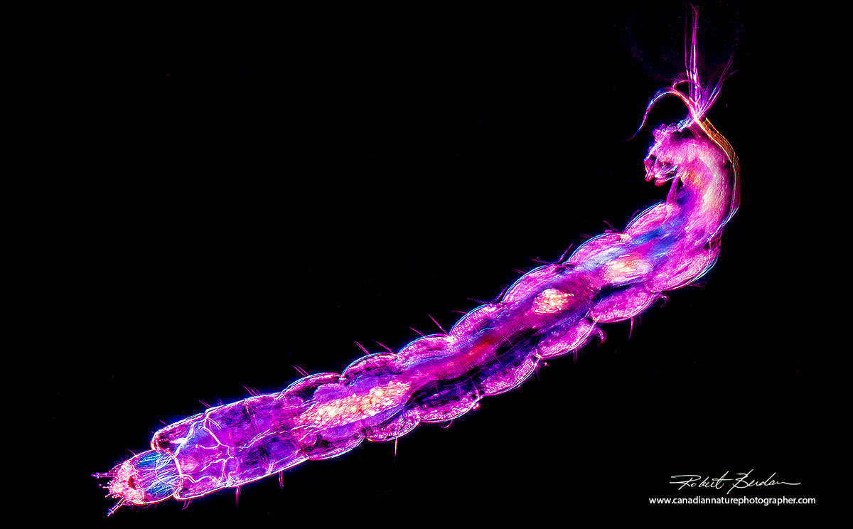
Arthropod larvae (Biting Midge Larva) viewed in Polarized light microscopy. Birefringent components appear coloured in polarized light. Below the photomicrograph shows a single segment from the arthropod above and a branch like structure which is a Trachea that forms part of the respiratory system of insects. About 40X

Single segment from the biting midge larvae above showing a branched trachea used by the organism to breath. The blue and red colours are muscles that have proteins (myosin and actin) aligned and these rotate the polarized light to cause interference and hence the colours you see above. About 100X
Chironomid larvae develop into small flies that resemble mosquitoes but they don't bite. The larvae can be several millimeters long. Their bodies are segmented, include a few bristles and they have sharp jaws and a breathing apparatus on the posterior end. Combined Darkfield and Rheinberg lighting about 40X.
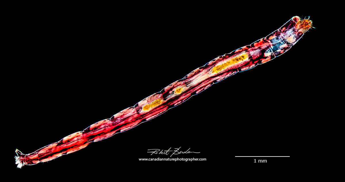
Chironomid larvae that was blood red, the red colour is from hemoglobin which helps larger animals breath in poorly oxygenated water. Panorama stitched from several images.

Chironomid larvae by polarized light microscopy showing the longitudinal muscles inside its segmented tube shaped body. Chironomids can be seen in a jar of pond water - as kids we used to call them "wigglers". Chironomids are also used in Fly Fishing as bait.
Chaetogaster (Annelida) worms live in freshwater and feed on small crustaceans and insect larvae. Their mouths are large, they are generally colourless and possess small hairs or bristles, called setae or chaetae located along the outside of the body. There are more then 800 species in North America. The mouth is at the right side of the picture. Darkfield microscopy.

Chaetogaster by Differential Interference microscopy (DIC). About 100X.
Hydra
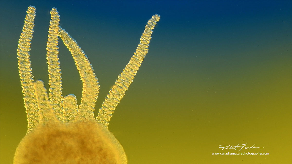
Hydra are common in ponds around Calgary in late July and August in my experience. The best place to collect them is in the Sibbald creek fish pond in Kananaskis. They are found attached to water plants so be sure to collect some plants with the pond water. They may be up to 1-5 mm long with several tentacles that have stinging cells that capture and paralyze their prey which often include copepods and water-fleas (see my previous article on Hydra). Their stinging cells, unlike those of jelly fish, will not harm you and can't penetrate your skin.
Hydra are of interest to researchers because these anaimals can regenerate quickly and as far as we can tell they show no signs of aging unlike most other animals. Some Hydra have a symbiotic relationship with green algae which live inside their cells - the green hydra.

Brown hydra by polarized light microscopy, about 50X. The cnidoblasts or stinging cells are birefringent and appear like tiny Christmas lights.

Above closer view of a hydra and the tentacles by polarized light microscopy 150X.
Close up view of a single tentacle from the hydra above. You can see the cell membranes, some cnidoblasts or stinging cells. This organism has an outer and inner layer of cells separated by non-cellular mesoglea. 400X Darkfield microscopy.
Hydra tentacles are used to capture prey like water fleas and copepods - Rheinberg lighting about 100X
Cladocerans
The Cladocera are a group of small crustaceans collectively called water fleas. There are over 650 species. They provide food for small fish and are currently being studied to see what the effects of global warming might be on them. Most range in size from about 0.2 mm to 3 mm. All have a distinct head and body covered by a fold of the cuticle. They are an important food source for fish and other small invertebrates like Hydra.

Cladoceran possibly Alona sp. 200X Darkfield microscopy and Rheinberg lighting. See my article on photographing water fleas.

Waterflea Simocephalus sp 50X viewed by polarized light microscopy, a full wave plate creates the pink background colour in polarized light. Muscles appear blue and yellow in polarized light. Three eggs can be seen on the left dorsal side and a large compound eye at the top of the organism.
Water Mites
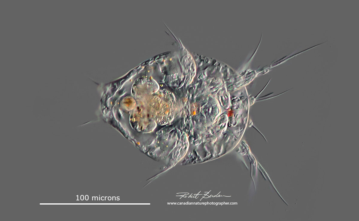
Water mites belong to the class Arachinda which includes the spiders and are in the subclass Acari. A few are aquatic (Hydrachinidia). Adults are needed for species identification. Water mites feature three active stages: larva, deutonymph and adult. Larva have only three pairs of legs shown below. The free living larva attaches to an immature aquatic insect and enters into a parasitic stage where if feeds on the host (Clifford, 1991 - Aquatic Invertebrates of Alberta - University of Alberta Press). The ones shown below were red in colour and have 2 prominent eyes. These one were less then a mm in size so a magnifying loupe or stereo microscope is required to see them.

Water mite 100X Darkfield microscopy.


Above water mite DIC microscopy (blue background) and below Darkfield microscopy about 40X
Copepods
Copepods are a group of small crustaceans found in most freshwater and saltwater habitats. They can be easily seen with a stereo microscope. Females often have two egg sacs attached to their posterior end. When the eggs hatch they form a larva stage called a nauplius which molts several times before becoming an adult. The adults have a single eye on their anterior end and for this reason are sometimes referred to as a cyclops.
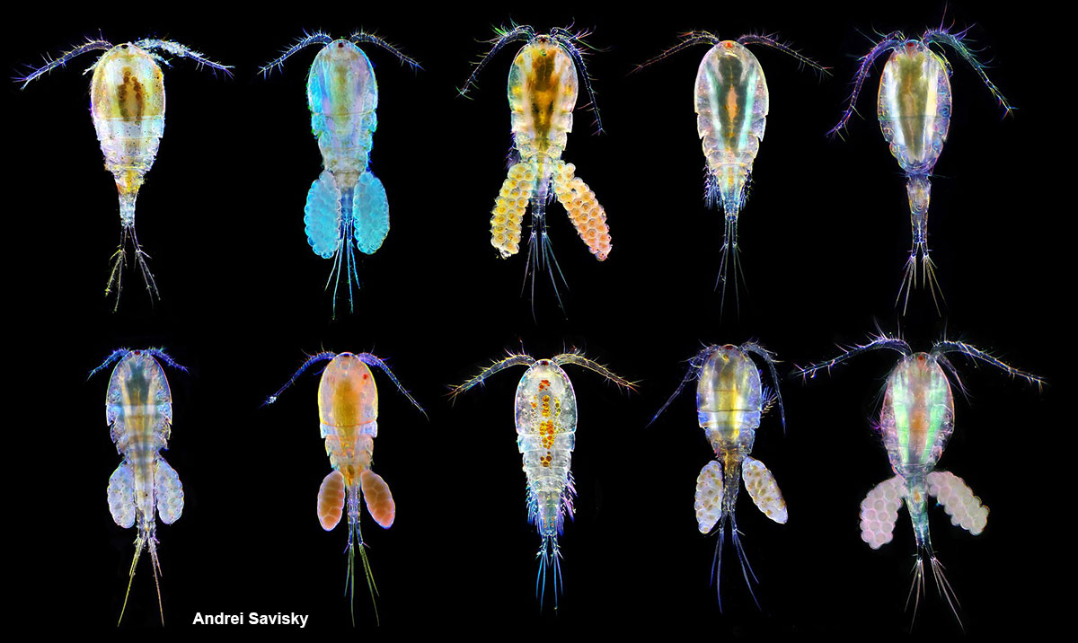
Above - photomicrography of an arrangement of adult copepods, photographed with combined Darkfield and polarization microscopy - Wikipedia commons by Andrei Savisky. About 50X.The adults can be 1-2 mm long and are an important food source for small fish.
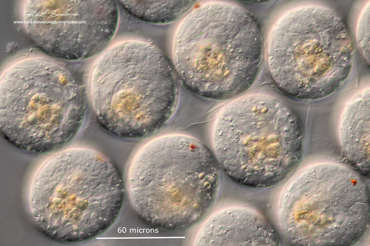
High magnification view of the eggs attached to a female copepod. You can see the red eye spot in some of the eggs.

View of copepod eggs using Polarization microscopy.
When copepod eggs hatch they form a nauplius larva that undergoes several molts before becoming an adult. They are very common in pond water and also in the ocean. Note the single red eye. Focus stack of images. DIC microscopy.
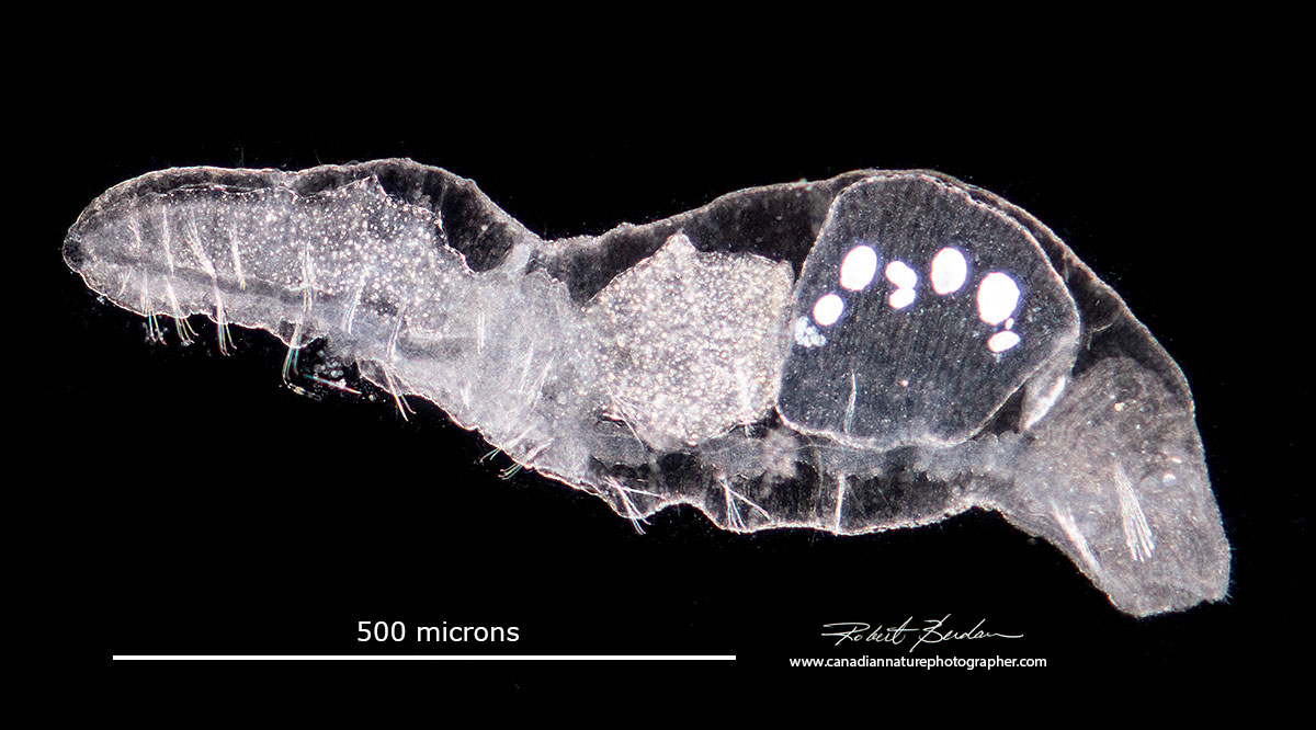
Gastrotich

Gastrotrichs are sometimes called hairybacks because of the spines on their body. Others refer to them as "cats" of the micro-world because of whiskers on their head. They vary in size from about 60-300 microns in length. Often they are found among plants and they can swim fairly fast making them challeging to photograph. The majority of gastrotrichs feed on detritus. Freshwater species reproduce parthenogenetically (without fertilization). They have short lifespans of only a few days and are multicellular. The one shown above is carrying a large egg inside. Single framegrab from a 2K video, DIC microscopy.
Rotifers
Rotifers are small multicellular micro-organisms common in pond water, water in moss, bird baths, and rain gutters. It's estimated there are about 2000 species of rotifers, most live in freshwater. They can occur in numbers of up to 5000 per litre. They play a role in nutrient recycling, they are used in testing water toxicity, and are also used to feed fish larva and even are sold as fish food in some pet stores (e.g. Brachionus plicatilis). To learn more about rotifers and see more pictures of these alien-like animals see my article on rotifers on this site.

Squatinella rostrum (Rotifera: Monogononta, Ploima, Lepadellidae) note 2 red eyes on the side of the head. DIC microscopy.

Bedelloid rotifer found in from water collected from a sidewalk puddle.
Dinoflagellates
Dinoflagellates are single celled protozoa that have hard shelled plates (theca made of cellulose). They posses one or two flagella they use to swim and sweep food into their mouth. They often appear to spin as they swim when viewed with a microscope. Dinoflagellates are considered to be protists, with their own division, Dinoflagellata. They are found in both marine and freshwater. Their are about 220 freshwater species. A bloom of certain dinoflagellates can result in a visible colouration of the water, colloquially known as a red tide, which can cause shellfish poisoning if humans eat contaminated shellfish. Some dinoflagellates also exhibit bioluminescence—primarily emitting blue-green light. I have seen bioluminescence dinoflagellates off the coast of BC in marine environments but not in freshwater.
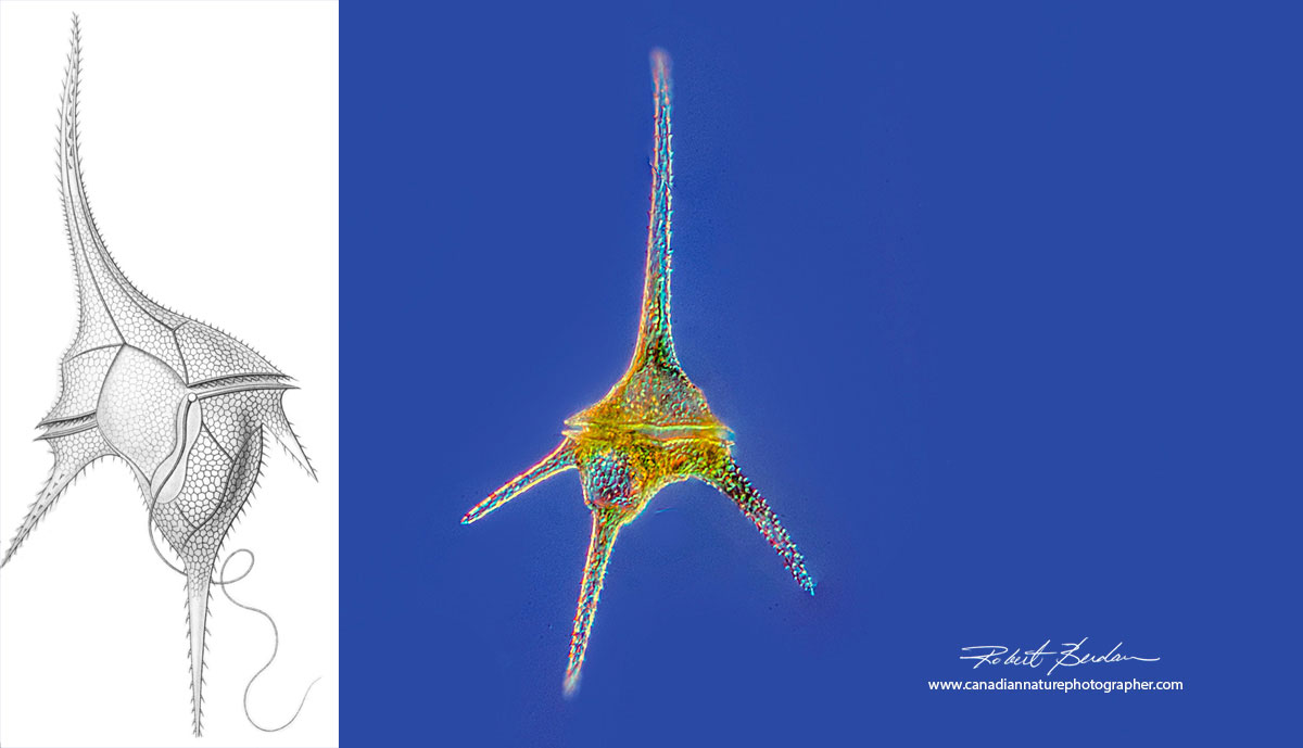
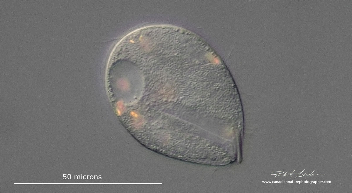
Above Ceratium hirundinella a common dinoflagellate in ponds and lakes in Alberta. On the left is drawing by Ernst Haeckel (1834–1919) - from Wikipedia. The photograph was taken using DIC microscopy and a focus stack of 15 images 400X.
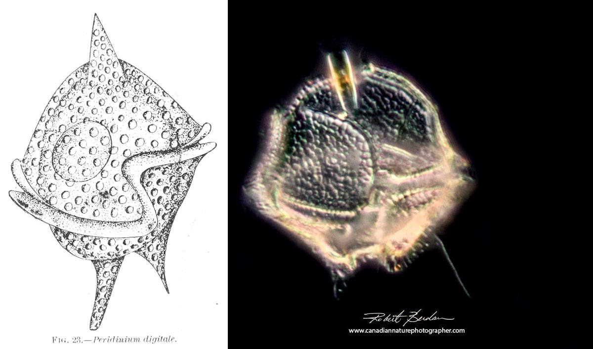
Above freshwater dinoflagellate - Left drawing from Wikipedia and on the right is a focus stack of a similar dinoflagellate from fresh water. Darkfield microscopy about 400X.

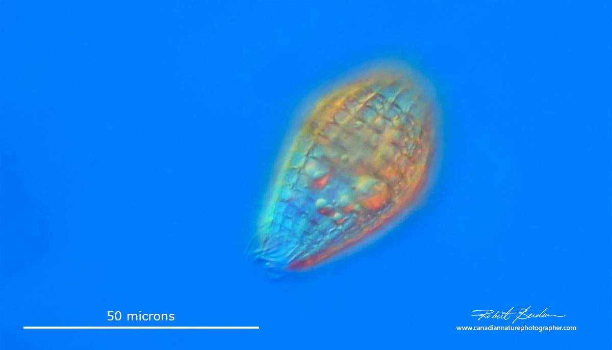
Peridinium sp of Dinoflagellate. Focus stack. Their shape makes it difficult to get the entire subject in focus. They often appear gold in colour and their flagella allow them to spin in the water.
Ciliates
Ciliates are a group of protists characterized by cilia and two nuclei. There maybe as many as 30,000 species of ciliates - many still to be discovered. While I have seen a number of unusual ciliates identifying them to species often requires silver staining, DNA analysis and considerable expertise. Below I include a few unusual species I have come across.

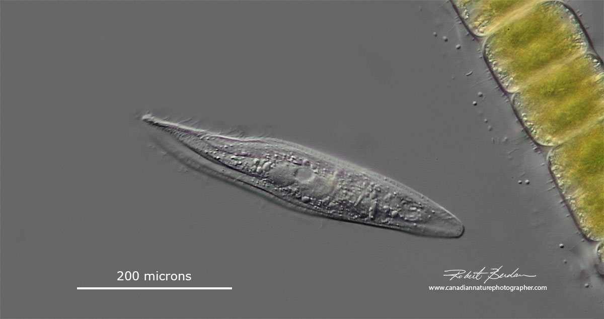
Lacrymaria olor is a species of ciliates, typically 100 micrometres (0.10 mm) long, that is found in freshwater ponds. It's neck constantly probes the environment and it can collapse or extend its neck as shown below. This ciliate is notable for its ability to extend the anterior end of the cell up to 7 times its body length, and manipulate it in many directions — even around obstacles — in order to capture food. DIC microscopy from a single video frame.
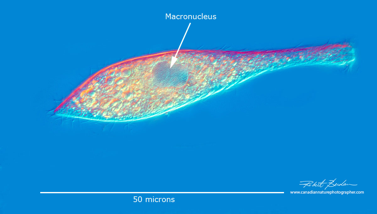
This ciliate has two macronuclei and a single micronucleus. Its entire cell body is covered with cilia arranged in spirals. DIC microscopy.
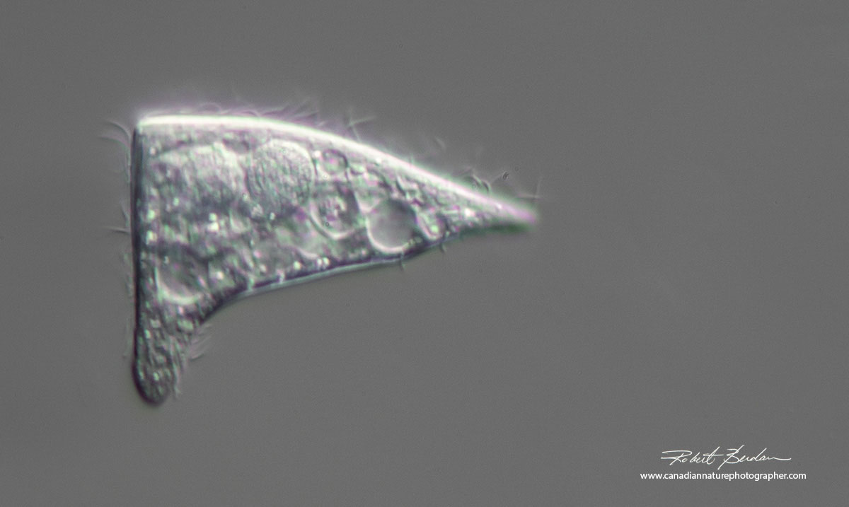
Triangular shaped ciliate I found in pond water. 400X DIC microscopy. Stokesia vernalis? alternatively this could be a damaged or deformed ciliate though I saw several.
Lembadion lucens? DIC microscopy.

This ciliate had a deep oral groove. Polarized light microscopy. Lembadion lucens?
Ciliate Coleps sp has a barrel-shaped body and test made of biomineralized plates. Usually they are under 100 microns long and have a distinct cage-like plates that contain both organic and inorganic components mostly of calcium carbonate. They feed on bacteria, algae and flagellates. DIC microscopy.
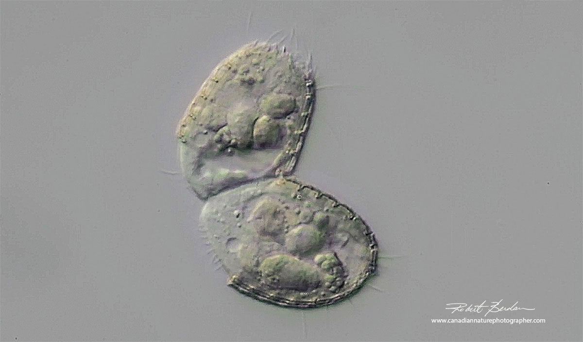
Ciliate Coleps dividing DIC microscopy
Puddle Ciliates
A friend from Calgary, Josh Grosse brought over some samples of water he collected in puddles where he had seen some unusual ciliates.
The first puddle ciliate is shown below. It moved rapidly and then would stop for a second or two and spread some spines before moving on again. He identified at least 9 different ciliates in the samples whereas I was only able to see 3 or 4 different ones.

Hastatella radians is 40-60 micron long ciliate with distinct spines. DIC microscopy

This puddle ciliate had a short proboscis.400X DIC microscopy
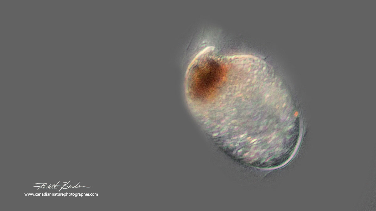
Puddle ciliate with a very short proboscis and brown pigmentation. DIC microscopy.
Ciliates from Pond water
Litonotus fasciola has 2 macronuclei visible - DIC microscopy

Peritrich stalked ciliate - the stalk was non-contractile and unbranched - Epistylid. DIC microscopy. 400X.
Flagellates & Algae
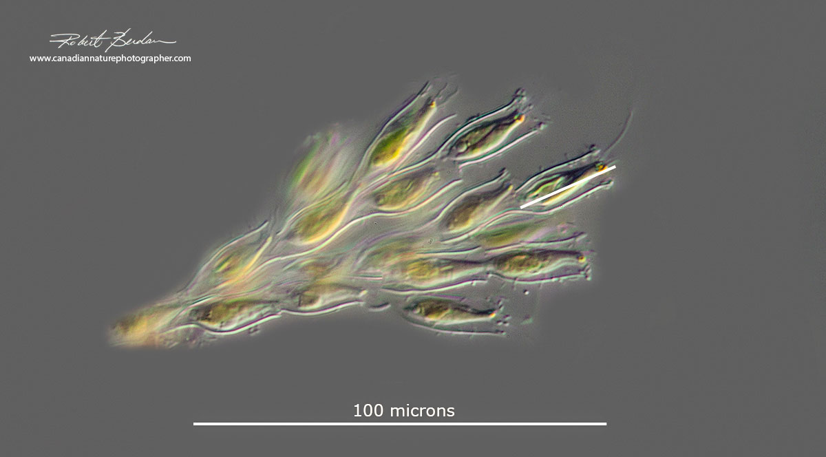
Dinobryon is a form of microscopic algae found in ponds. I often see just the empty loricas and I am still working on getting some better pictures. They are common in freshwater lakes and ponds. There are 22 genera and 37 different species. They are mixotrophs meaning they can get energy from photosynthesis and through phagotrophy of bacteria. They have a single long flagellum and red eye-spots and the organisms are protected in a vase-shaped lorica. For more information see Science Direct.
Volvox
Volvox belongs to the chlorophyte green algae. They live in freshwater and can form spherical colonies of up to about 50,000 cells. To see more pictures and learn more about Volvox see my other article on this site.

Volvox photographed with Darkfield microscopy, smaller cells with flagella can be seen with red eye spots that comprise this sphere. The sphere often rotates as the organism swims. The cells divide asynchronously allowing the organism to remain afloat during cell division. Daughter colonies often form inside and later break out of the interior.
Algae
Blue-green algae are named after their colour and are also known as cyanobacteria. This group of organisms like bacteria lack a nuclear membrane, the DNA is usually circular and floats directly in the cytoplasm. These are among the earliest known organisms on the planet and are responsible for forming Earth's early oxygen. Fossils of some species of cyanobacteria have been dated to 3.5 billion years old. They are common in stagnant water, hot-pools in Banff National Park, bird-baths, and ponds. Some species can produce toxins that can kill animals that drink the water including cattle and ducks. Their cyanotoxins can also accumulate in shellfish causing poisoning to those that eat the shellfish.

Oscillatoria is a common blue-green algae named after its oscillating movement - the tip of the trichome oscillates like a pendulum. The individual filaments appear to slide back and forth when viewed with a microscope. You can see distinct separate cell walls in the filament above and its blue-green colour. Each filament of oscillatoria consists of trichome which is made up of rows of cells. Oscillatoria are the subject of research into the natural production of butylated hydroxytoluene (BHT), an antioxidant, food additive and industrial chemical - Wikipedia. DIC microscopy.
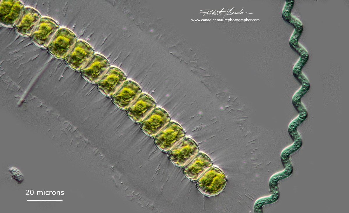
Above is a green algae (left) Hyalotheca (Desmidiaceae) and blue-green algae species on the right (Arthrospira sp) that forms single spiral shaped filaments. This filament was non-motile and about 16 species are known. For a recent report on the characterization of Arthrospira and Limnospira species see Nowicka-Krawczyka (2019) Scientific Reports Vol 9, Article number: 694. Hyalotheca posses a wide mucilage sheath with fibrils extending from the cells walls.
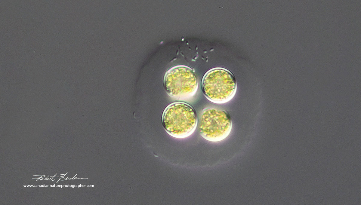
Green algae - possibly Tetrabaena socialis encased within a mucilage sheath. These algae are of interest in understanding how multicellularity came about through evolution - see Solving the puzzle of multicellarity. 400X DIC microscopy. Some researchers consider this algae as Gonium sociale also see Wikipedia.

Yamagishiella unicocca or Pandorina charkowiensis is colonial green algae in the family of Volvocaceae. The cells are encased in a thin mucous sheath. DIC microscopy (it has also been suggested to me that this algae might be Dictyosphaerium or Botryococcus).
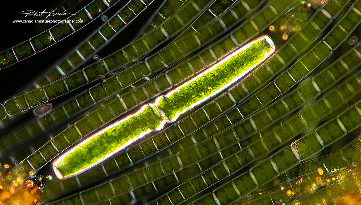
Pleurotaenium sp is a desmid (center) viewed by polarized light microscopy. These desmids can be up to 1-3 mm long and visible even with a stereo microscope. They are long, cylindrical and have a rough thickening in the central area where the two semicells join. They are common in pond water throughout the summer. Polarized light microscopy.The background filamentous algae are Microspora sp.

Pleurotaenium sp via polarized light microscopy, the pink background is created by using a full retardation wave plate with polarizing filters.
Heliozoan
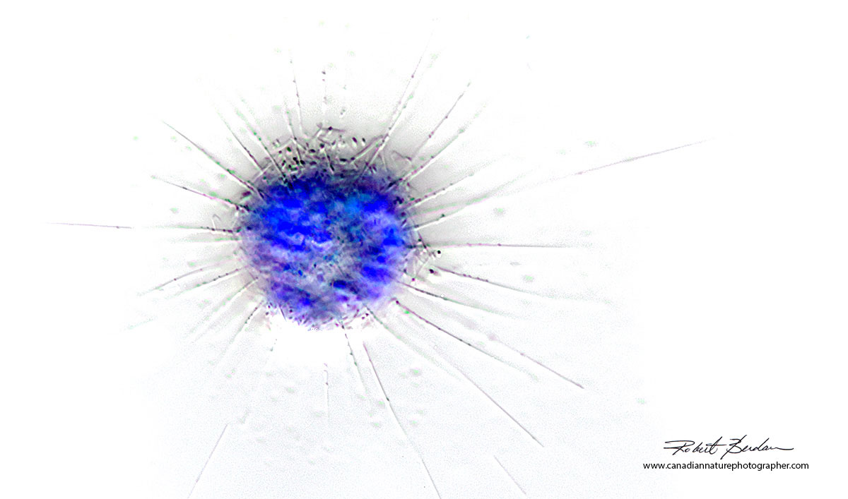
Above is a Heliozoan (Heliophyra rotunda?)- also called sun-animalcules, with arms called axopodia that it uses to catch prey and absorb the contents of their prey. The axopodia are microtubule-supported projections from the amoeboid cell body, and are variously used for capturing food, sensation, movement, and attachment. I am not able to identify the species in this photograph which I inverted the colour. Darkfield microscopy 400X.
Unknown Pond Cyst

This photo shows what seems to be a single cell encased within a mucilage sheath. The green and yellow colours suggest it is an algae and there is a single red eye. It was non-motile and could be resting or cyst stage. Cysts are common in pond water e.g. rotifers and this is a method that permits many pond microorganisms to survive winter or when the pond drys up in summer.
If you are curious about what might live in ponds, find a pond near you, bring a few clear pastic jars, some nets and a large plastic Turkey baster ($3 from Walmart) which acts like a giant eye dropper to take water and mud samples. A small lens loupe or jewlery loupe will help you see the smaller organisms in your jar and one can be purchased for under $20.
Notes on Photography
All pictures were taken with a Zeiss Axioscope microscope, Nikon D500 camera shooting RAW files or video at ISO 100 - 200. The camera was attached to a Laptop (Alienware) via a USB cord and the laptop was running Digicam control software (free). I photographed in Liveview mode and processed the images in Adobe Photoshop. For some photos I used focus stacking to achieve a greater depth of field. Dimensions were determined with an eyepiece micrometer calibrated to a slide micrometer. All microorganisms were collected from around Calgary in puddles, ponds or small lakes (Sibbald Creek pond, Winchell Lake).
Acknowledgements:
I thank Josh Grosse from Calgary for bringing over samples of water he collected from puddles and for identifying the ciliates in those samples. Also for his suggestions on the identification of Arthrospira and Hyalotheca and the Biting Midgle larvae.
Note on identification of Ciliates. The identify of some ciliates requires research experts and may involve silver staining or DNA analysis - the identity of the organisms in these pictures are based on keys that I have in my library and in some cases represent my best guess.
References and other web sites:
1. Microorganisms in a Pond
2. It came from the Pond - ciliates - the web master is a ciliate expert
3. Microscopic Pond life
4. Observing life from a pond - Microscopes for schools
5. Aquatic Animal Identification Guide - Kananaskis country, Alberta - PDF
6. W. Foissner - World expert on Ciliates and other pond life - download his free PDF books
7. Harmut Bick - Ciliated Protozoa (1972) World Health Organization - Key free Ebook PDF
Authors Biography & Contact Information
 Bio: Robert Berdan is a professional nature photographer living in Calgary, AB specializing in nature, wildlife and science photography. Robert retired from Cell\Neurobiology research to take up photography full time years ago. Robert offers photo guiding and private instruction in all aspects of nature photography and Adobe Photoshop training - including photomicrography, macrophotography.
Related Microscopy Articles by Robert Berdan on this web site
1. Photomicrography of Hydra - a model for studying regeneration and aging
2. Photographing Daphnia
3. Photographing Gastrotrichs
4. Photographing Rotifers
5. Photographing Ciliates
6. Photographing Stentors - A Large Unicellular Protozoan (ciliate) living in Freshwater
7. How to Collect and Photograph Water Bears (Tardigrades).
8. Tips on How to take Better pictures with a Microscope
9. Microscopic Pond Organisms from Silver Springs Calgary
10. Microscopic Life in Ponds and Rainwater - Pond Scum I
11. Photographing Microscopic Plant and Animal Life - Pond Scum II
12. Photomicrography and Video of Protozoa, Volvox and Rotifers
13. Home Microscopy Laboratory for Photomicrography
14. The Art & Science of Photomicrography with Polarized Light
15. Photographing Through a Microscope Photomicrography - Inner Space
16. Focus Stacking comparing Photoshop, Helicon Focus and Zerene
17. Rheinberg Filters for Photomicrography
18. Scanning Electron Microscopy - Photography
19. Photomicrographs of Diatoms from 1877 by John T. Redmayne
Email at: rberdan@scienceandart.org
Web site: www.canadiannaturephotographer.com
Phone: MST 9am -7 pm (403) 247-2457.
Click on the buttons below and share this site with your friends