
The Micro-Universe - Microscopic Life
by Dr. Robert Berdan
August 30, 2019
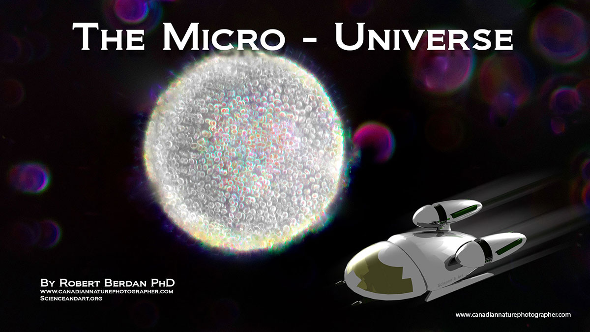
Above is a glimpse of pond water magnified 400X. The sun-like object is Volvox a colonial form of algae made up of thousands of individual cells. The purple objects are out of focus protists. I constructed the space ship in a 3D program and it is meant to be symbolic of a light microscope. The Micro-universe is all around us and contains thousands of alien-like creatures some of which I show below. Darkfield microscopy about 200X
Introduction
I define the Micro-Universe to include organisms from about 2 mm in size down to 0.0002 mm (2000 to 0.2 microns where a micron = 1\1000 of mm). Micro-organisms are easy to observe with a stereo or light microscope. Anyone can begin to explore the Micro-universe starting with a simple magnifying lens. Microscopes are better of course and the cost of an entry level light microscope starts around $75. Toy microscopes are cheaper and I started with one, but I think they are only suitable for young children. There are also paper microscopes that magnify 200X or more available for only a couple of dollars - see below.
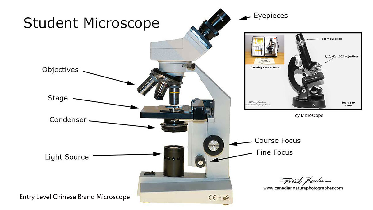
Above is a Chinese brand microscope I purchased for $100 used on Kijiji. My first toy microscope I received for Christmas in Grade 6 is shown in the inset photo and cost $29 from Sears. Frankly it wasn't very good, but it was good enough to see pond organisms and it inspired me to get something better 2 years later and the optics are better than the $2 paper microscopes shown below. There are also USB microscopes, I haven't used one, but I would suggest getting a stereo-microscope instead. Also if you have a flatbed scanner at home - it can function as low power microscope to get started - see my article.
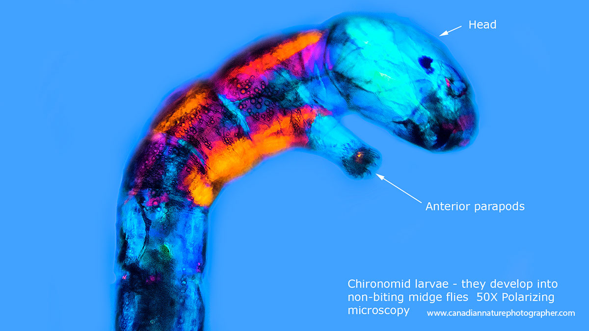
Chironomids are insect larvae that live in ponds and metamorphosize into small flies. 50X Polarizing microscopy.
The goal of this article is to give you a glimpse into the profound and fascinating micro-world. You don't have to visit another planet to see alien-like creatures. Some of these micro-organisms build intricate "houses" made of glass that are used in tooth paste and to filter beer. Some microorganisms cause disease and others (Tardigrades) may one day help us visit distant planets.
Multimedia Presentation on the Micro-Universe Available in Calgary
The pictures shown in this article are just a few images along with some movies of this amazing world. I have created a multimedia presentation that I would be happy to present to your group, club, or school near Calgary. My goal is to promote microscopy, photomicrography and inspire others to explore the micro-world. Microorganisms can be found everywhere: in your backyard, bird bath, ditch, pond, lake or stream. Even puddles that form on the sidewalk after a rainfall. If you are interested in having me present contact me rberdan@scienceandart.org.
Brief History of the Microscope
Hans and Zaccharius Janssen in the Netherlands are believed to have built the first compound light microscope between 1590-1610, while Galileo created the spyglass (10X telescope) in Italy in 1608. A Dutchman Antonie van Leeuwenhoek used single-lensed microscopes of his own construction to discover protists (The Protista are a kingdom of simple eukaryotic organisms, composed of a single cells or a colony of similar cells). Robert Hooke an Englishman used a two and three lens microscope made by Christopher Cock in London and with it he discovered the first cells in cork and published his findings in 1665 in Micrographia. You can download a free copy of Micrographia as a PDF if interested. His drawings include insects and snowflakes.

Images courtesy of Wikipedia

Check out the web site (www.foldscope.com) to learn how you can purchase paper microscopes for a few dollars. These paper microscopes were designed for those that could not afford better quality microscopes and for children. In my experience the image quality of these scopes is not great, but for a few dollars it might inspire some kids to become more interested in science and the micro-universe.
Microscopes are routinely used in medicine, research, pharmaceutical industries, health laboratories, forensic science labs, manufacturing, electronics inspection, engineering, metallurgy, microbiology, geology, surgery, chemistry, physics, botany, zoology, agriculture research, veterinary science, museums, art restoration, aquariums, fisheries, jewellers, environmental science, beer breweries and wineries. A microscope is one of the most important tools used in science and medicine. The microscope can also show us a things that most of us will never see and it has never been easier or cheaper to own one.
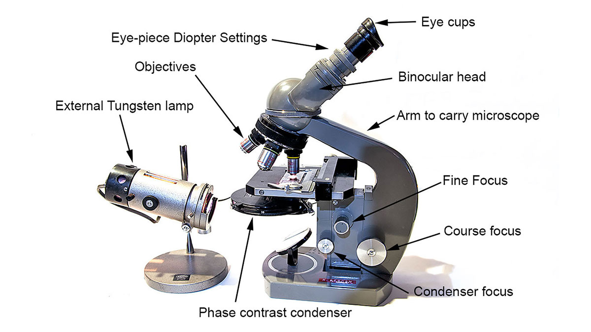
Above my first professional microscope (Olympus E) that cost me $300 back in the early 1970's - I purchased this scope after graduating from grade 8 with the help of my parents. Used versions of this scope come up on E-bay and other auctions for $75-$300 depending on the condition and accessories included. New Chinese microscopes can be purchased for $50-$300 that are excellent to start studying micro-organisms. They magnify up to 1000X - good enough to see bacteria and most other micro-organisms. A good place to purchase a new microscope is AmScope - these are Chinese made scopes and the quality to cost ratio is very high and I have been happy with their service. For tips on buying a microscope see my article on this web site.
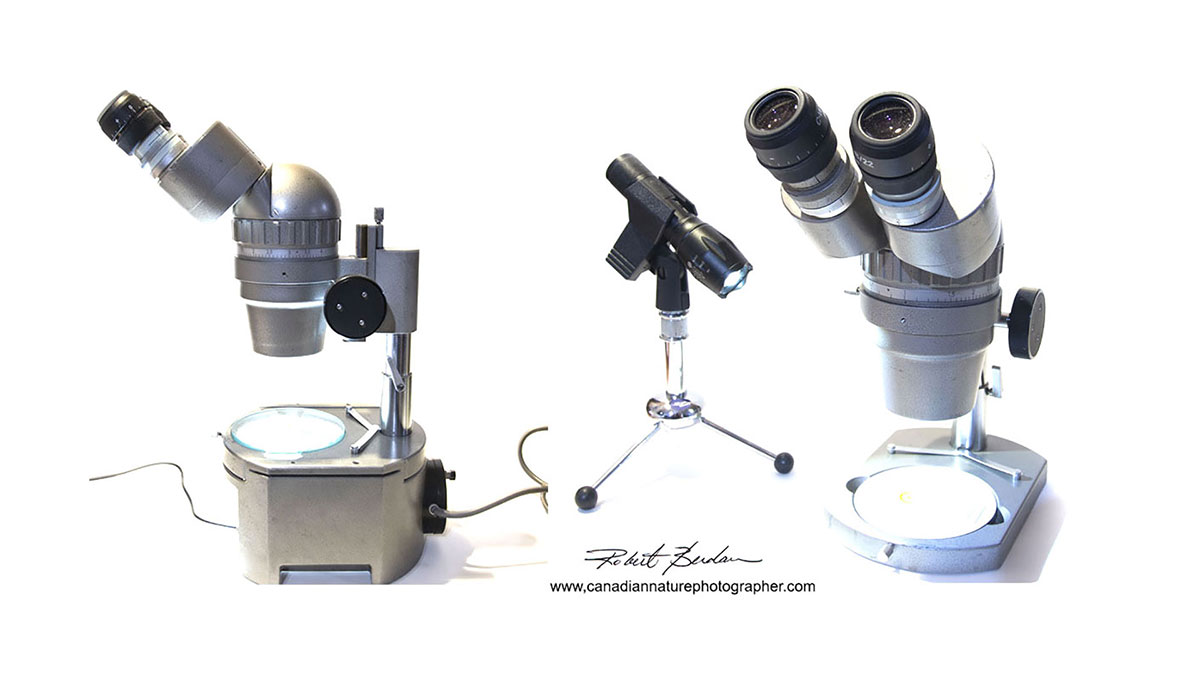
Above is an Olympus zoom stereo-microscope that magnifies from about 5-40X. These scopes are excellent for young children (9 years old and up) because they are easy to use, require no glass slides, and only a simple light source. I have attached an LED flash light ($20) to a microphone stand ($10) to provide a bright light source. Objects appear in 3D and this scope is great for studying plants, arthropods, lichen, rocks, fossils - even coins, stamps and jewelry. I would recommend a stereoscope as a first microscope for anyone especially kids.

Above is a picture of a painted lady butterfly from Alberta magnified 36X. This image was focus stacked to create a greater depth of field. Many insects appear alien-like and some of the wasps and beetles can have large jaws which can make them look scary.

Above is a common mosquito resting on human skin. I used a 100 mm macro lens to the take the photo that is magnified about 8X. The proboscis which the mosquito uses to suck blood from its host is shown below further magnified using a light and scanning electron microscope.

Mosquito proboscis on left viewed with a bright field light microscope, and on the right with a scanning electron microscope (false coloured) about 400X.
Insects in general make great subjects for studying with either a stereo or light microscope. Below are some photos of an aphid I photographed with a Scanning electron microscope and false coloured them (electron microscopes only provide images in black and white).
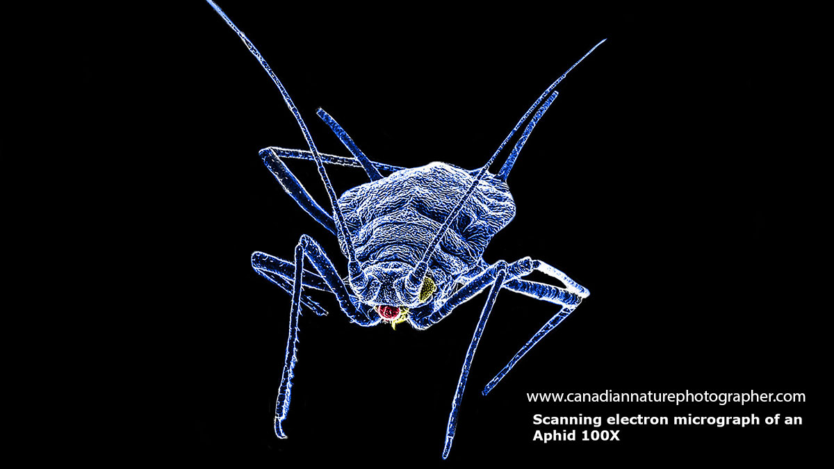
Aphid photographed with a scanning electron microscope (SEM) and false coloured.

Aphid about 400X SEM - note the compound eye (yellow) and mouth parts for sucking (green).

Close-up view of the Aphid's compound eye showing a single bacterium. SEM 10,000X.
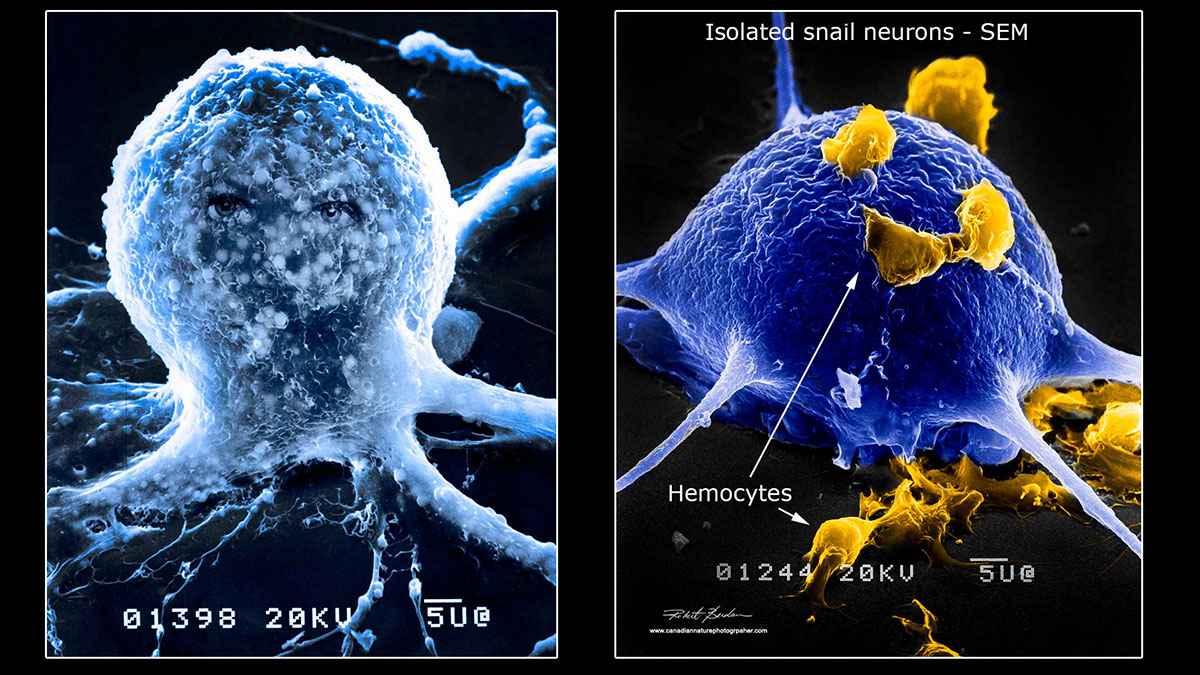
Above are single isolated neurons (brain cells) from a pond snail Helisoma trivolvis I used in my research to study nerve regeneration and synapse formation. Hemocytes are blood cells found in many invertebrates that have an immune function. SEM microscopy about 2000X.

Above scanning electron microscope from the University of Calgary. These microscopes are expensive (hundreds of thousands of dollars), but fortunately I can rent time on the microscope.

Above is a single membrane protein photographed at about 1,000,000X with a transmission electron microscope and then false coloured and enhanced to show 6 subunits surrounding a single pore. Gap junction channels allow your heart to beat synchronously and play other important roles between the cells in your organs and in nervous system.
Some of the most interesting microorganisms can be found in your backyard within moss, lichen, a bird bath, soil, or rain-water from your gutters. The best place to collect micro-organisms is from a pond, stream, ditch or lake. If you have on ocean nearby that is also a great source of microorganisms. Be sure to include some plants or algae as many microorganisms live on or around aquatic plants and algae. Some microorganisms live between sand grains. Nematodes shown below are particularly abundant in soil, just add water - see below.

My favourite place to collect microorganisms is in a pond. There are thousands of different species, including arthropods, snails, protists, diatoms, desmids, worms and algae. You can view larger insects and larvae by eye and with the help of a stereo-microscope discover smaller organisms or study details like their eyes and internal organs.

I find Amoeba frequently in water from my gutters and in ponds though be sure to collect some mud from the bottom of the pond and then later from the bottom of your jar. There are many different species of Amoeba some of them build "houses" or shells that they live in. 400X Differential interference microscopy (DIC). To learn more about amoeba visit Siemensma, F. J., Microworld, world of amoeboid organisms. World-wide electronic publication, Kortenhoef, the Netherlands.
Amoeba move slowly by a flowing of their protoplasm. Note that if you sample clean water, e.g. well water or tap water it should not show any microorganisms unless it's contaminated. Also you may have to concentrate the specimens, let them settle to the bottom over night. I also use an old centrifuge I purchased on Kijiji to concentrate specimens in a few minutes. You can also purchase a hand crank centrifuge on Amazon. Microcentrifuges can be also be purchased on Ebay for under $100, and the small plastic test tubes are available from Amazon.
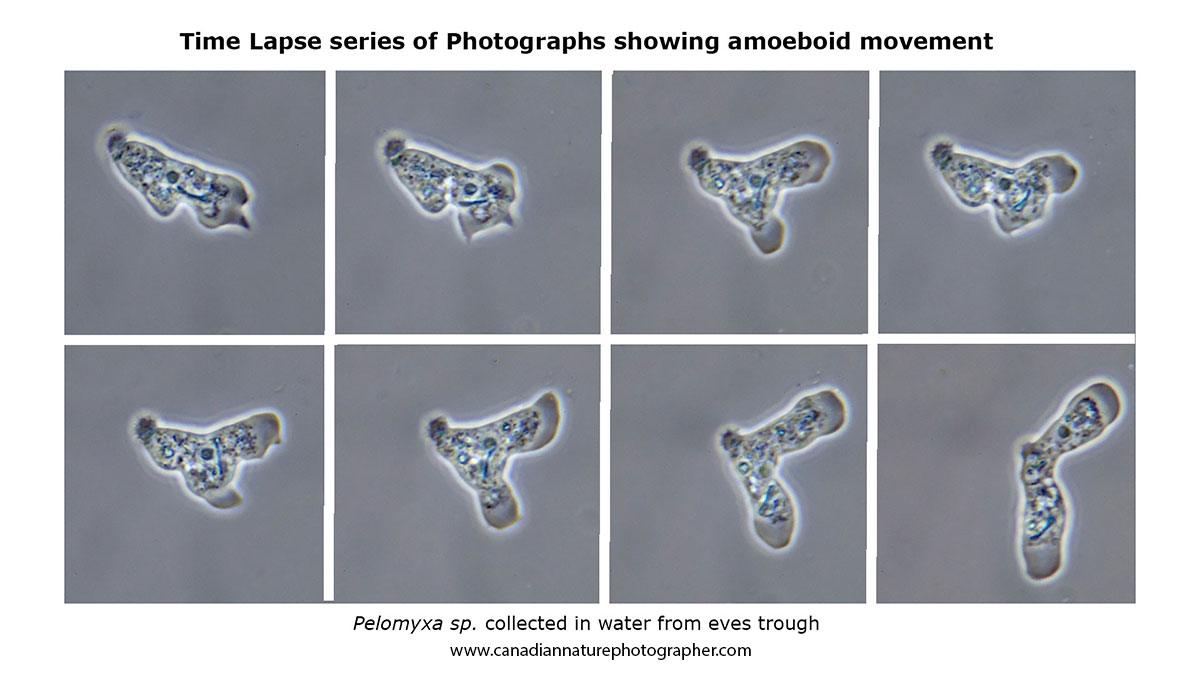
Above is a series of time-lapse photos showing how an amoeba changes shape as it moves over the microscope slide - 5 seconds between photographs. Phase contrast microscopy 400X. Pelomyxa have multiple cilia, and are anaerobic. The amoeba above has a spiny like-tail or uroid that suggested to me it might be Pelomyxa, but its identity is uncertain.
A very common microorganism found in pond water are ciliates. Ciliates have small hair-like projections called cilia which allow them to move and also sweep food like bacteria into their mouths. They also have two types of nuclei - a macronucleus and smaller micronuclei. Ciliates range in size from about 10 microns to 1000 microns (1 mm) or more and many of them can move quickly making them a challenge to photograph.
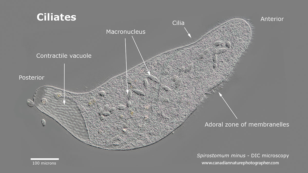
Spirostomum minus is a large common ciliate from pond water - it is 2-4X larger than paramecium shown below.
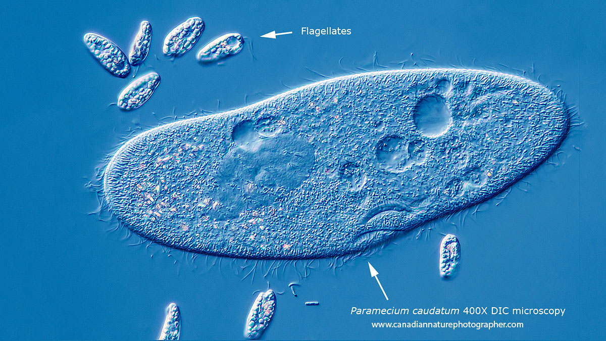
Above is a Paramecium caudatum surrounded by smaller flagellates. Paramecium are usually about a third the size of Spirostomum, but the higher magnification in this photo make the paramecium look similar in size. DIC microscopy.
Pond Organisms including Parameciium caudatum, Urocentrum turbo, Diatom and other flagellates. DIC microscopy. Watch on YouTube: https://www.youtube.com/watch?v=OwaPLjHOPVY
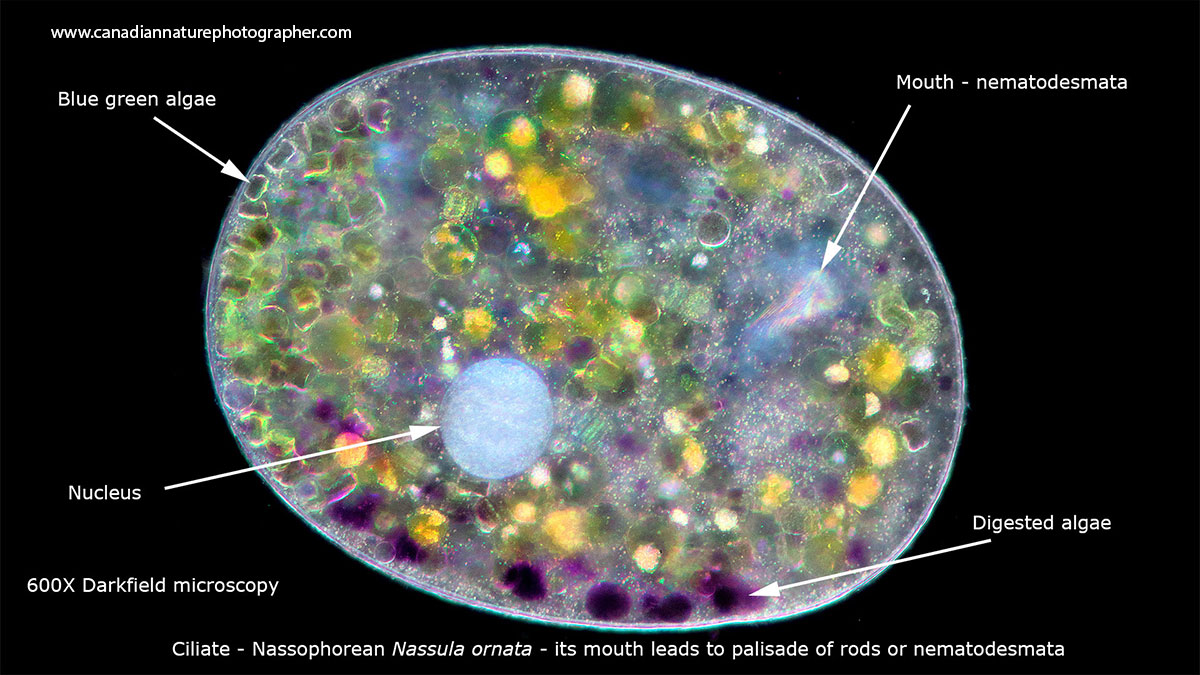
Nassophorea are a class of ciliates where their mouth leads to a curved cytopharynx supported by a palisade of rods. The one above has been feeding on blue green algae that turns purple as they are digested. Darkfield microscopy about 600X.
Volvox
Volvox is a colonial form of algae that can contain up to 50,000 cells. They often appear bright green in colour and rotate while they move around and can be several mm in diameter. They are made up of smaller single celled algae that are interconnected, each cell has 2 flagella which it uses to propel the organism. Reproduction involves the formation of daughter colonies that form inside the sphere that later break out and form a new colony. To learn more about Volvox see my article on Protozoa.
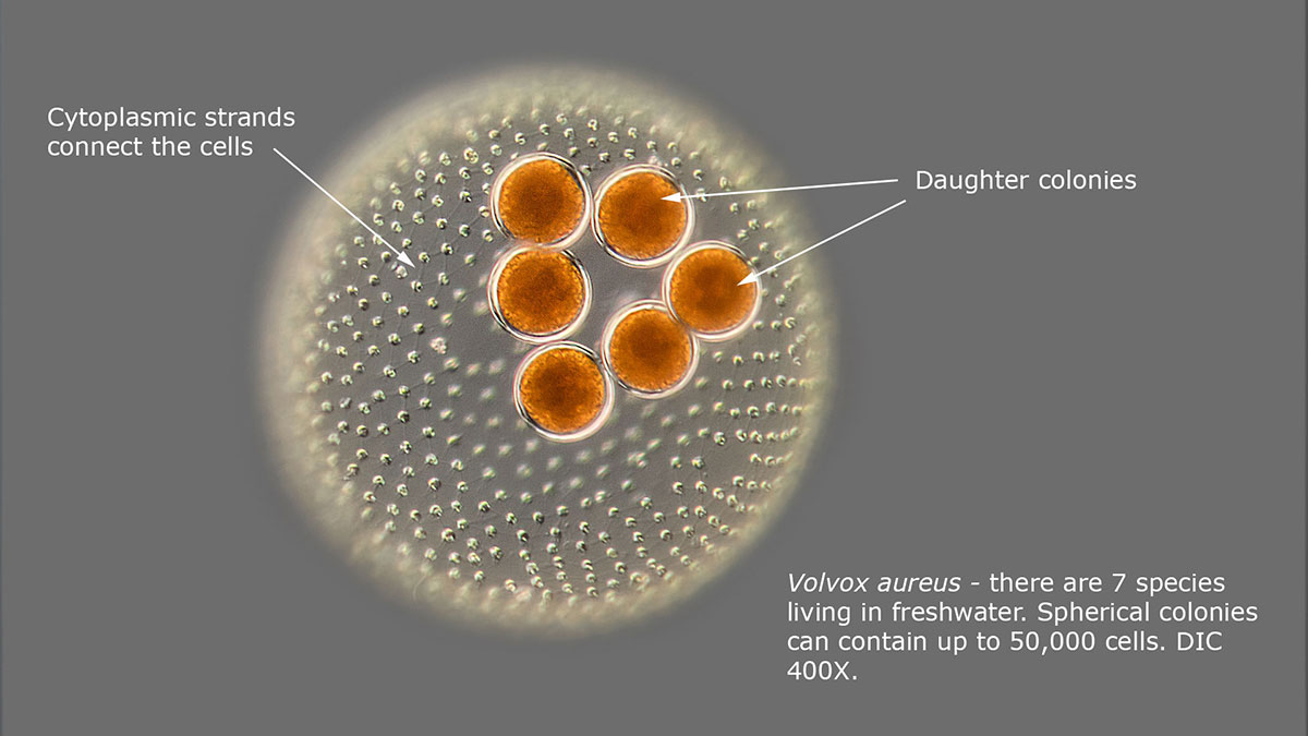
Volvox and daughter colonies, DIC microscopy

Higher magnification of Volvox showing the individual cells and their flagella. DIC microscopy.
Rotifers
Rotifers are microscopic sized animals made up of about 1000 cells. They are called rotifers because they often show one or two circular arrangements of cilia that spin like wheels which in turn sweep food and bacteria into their mouths and can also help propel them. Rotifers come in a variety of shapes and sizes, some like Rotaria below can telescope its body to several times its length. Many reproduce via parthenogenesis which is a form of reproduction without fertilization and some species of rotifers consist of only females. Rotifers also form cysts that can survive dessication when the pond dries up. Many species have prominent red eyes. For more about rotifers see my article on this site.

Euchlanis species of rotifer. The Trophi act as jaws to crush food and are also important taxonomically and assist in the identification of different species. DIC microscopy 400X.
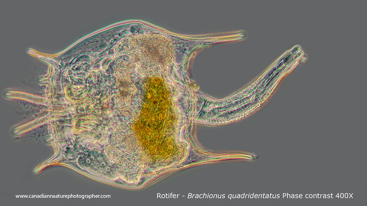
Above Brachionus quadridentatus. Phase contrast microscopy 400X.

Above Rotaria sp is able to extend its body 3X its length by extending in a telescopic manner. 100X DIC microscopy
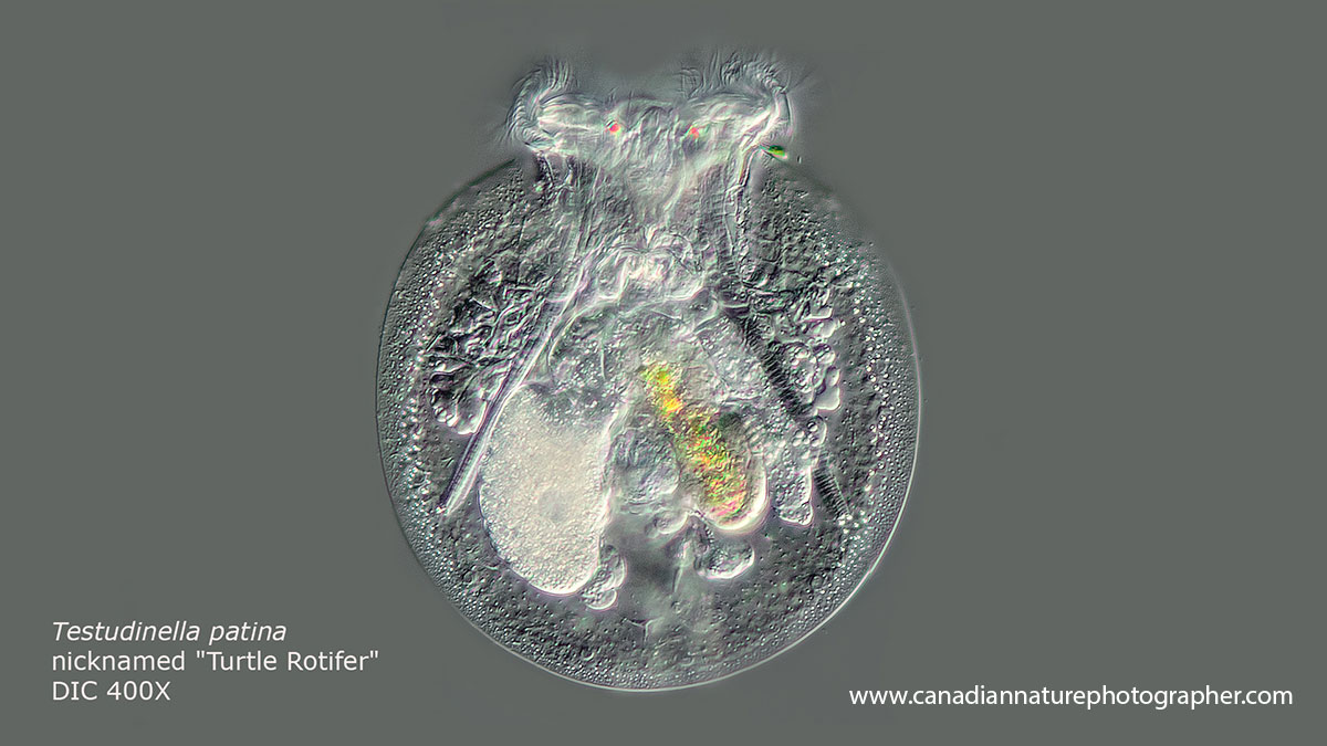
Testudinella patina also called the turtle rotifer, it is one of my favourites. It has a tail and two distinct red eyes at the top. DIC microscopy 400X.
Hydra
Hydra is a simple metazoan consisting of two cell layers including an outer ectoderm and inner endoderm separated by an acelluar mesoglea. The animal forms a flexible tube with long tentacles. The tentacles contain stinging cells (cnidoblasts) which are used to capture and paralyze prey which they then pull into their gastric cavity where it is digested. This animal is of interest to research scientists because of its incredible ability to regenerate and hydra do not appear to age. To learn more about hydra see my article on this site. Hydra are common in ponds often attached to algae and other water plants. A rich source of hydra around Calgary is the Sibbald creek fish pond in Kananaskis.

Above brown hydra feeding on a water flea. Brightfield microscopy about 40X.
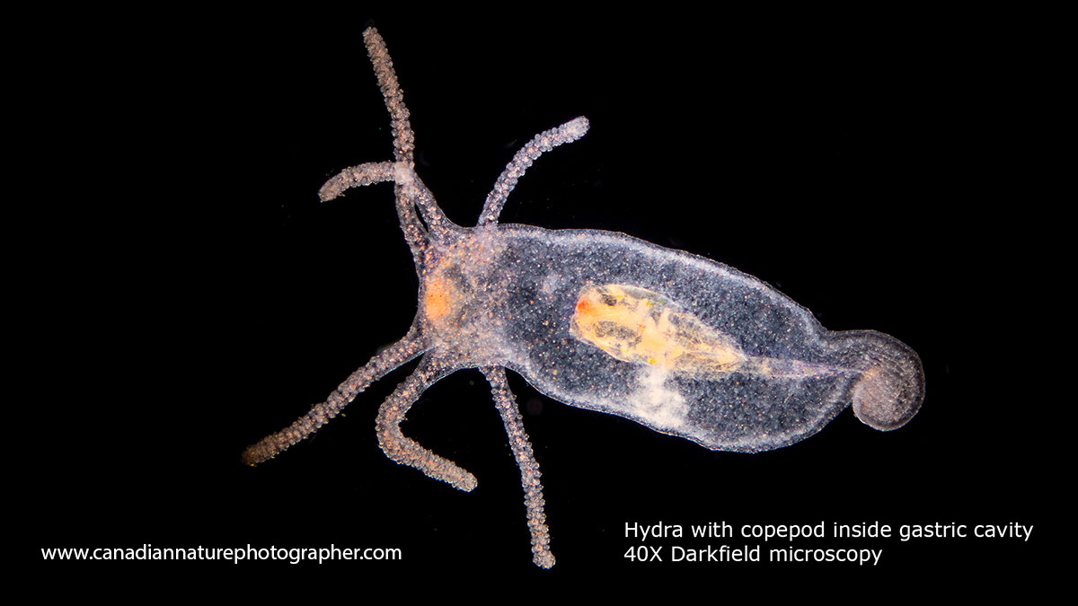
Above is a hydra that has swallowed a copepod which you can see in its gastric cavity. 40X Darkfield microscopy.
Diatoms
Diatoms are single cell organisms commonly found in streams and ponds. They can be a nuisance and form "rock snot" a brown slippery layer on rocks in the Bow river. However, under the microscope they appear as intricately shaped glass shells made of silica with ornate patterns and wide variety of shapes. Many of them are motile and move slowly over the substratum. The diatoms shown below have been cleaned of their internal contents which are normally golden brown in colour from their photosynthetic pigments. They are one of the most beautiful microorganisms and resemble jewels. Diatoms are used in silver polishes, as filters for beer and have many other uses. The best way to see or collect them is to scrape the slime off rocks found in water - I use an old tooth brush. In the 1800's some collectors would arrange the diatoms on microscope slides. See photomicrographs of Diatoms taken in 1877 by John Redmayne. To learn more about diatoms and get help identifying them see this web site diatoms.org and download guides from the net e.g. "A Treatise on Diatomaceae" by Henri Van Heurck (1896) with thousands of drawings - PDF.


Above photomicrographs of cleaned Diatoms taken with DIC microscopy. Live diatoms can be cleaned using household bleach which removes the internal contents and photosynthetic pigments. I created this arrangement of Diatoms using Photoshop.
Desmids
Desmids are algae that can also take on fascinating shapes, but their cell walls are made up of cellulose rather then silica. Below are just a couple of photomicrographs of these ornate specimens. They are found in pond water and can be collected easily around the edge of lakes and ponds by pushing a small lid into the moss around the body of water and collecting the fluid that fills the lid. Of course you can just collect water, but they seem to be more concentrated when I collect them this way. For instructions on how to collect desmids see this web site.

Above Pleurotaenium sp of desmid viewed by polarization microscopy 400X

Above is the desmid Micrasterias, which is fairly common in pond water. Darkfield microscopy.
Radiolarians
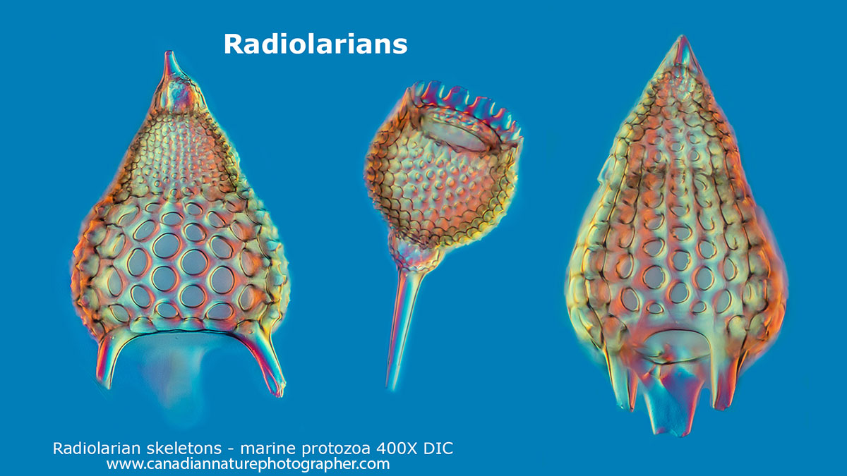
Radiolarians live in the ocean, however it is possible to buy prepared slides to view them. They are protozoa about 100-200 microns in size with elaborate mineral skeletons made of silica like the diatoms. They are important in geology and oil exploration. They were drawn and studied by Ernst Haeckel a German zoologist who's books and artwork are still being sold today as art. His most beautiful book is sold by Taschen but you can view some of his drawings for free in online e-books PDF. I first encountered radiolarians in a marine biology course I took in St. Andrews New Brunswick - Huntsman marine center while in 4th year University and I took the photo in black and white below with my Olympus E microscope.
Above Radiolarians from fossil samples from the Netherlands. Focus stacks, DIC microscopy.

Radiolarians from samples taken at the Huntsman marine center in New Brunswick, Canada. Darkfield microscopy
Water bears (Tardigrades)
Water bears are small 8 legged animals that live mostly in moss and lichen though they are also found in pond water and the ocean. They are referred to by their common name as Water bears or moss piglets. Most feed feed on plants they pierce with sharp stylets. A few Tardigrades are predatory feeding on nematodes, rotifers and other Tardigrades (e.g. Milenisum sp. below). These animals are about 0.2 to 0.5 mm, but some can grow up to about 1 mm. They do not cause disease and so far have no economic significance. However, they are unusual in that they survive dessication, X-rays, UV light, extreme temperatures and even in the vacuum of outer-space. They are common in the water-film within moss but when the moss dries up they form a tun stage where the exclude most of the water from their bodies until there is no biological activity. In this resting state they can survive for years and one researcher referred to them as time-travellers for this reason. I have found them almost every where I have looked.
I have at least three different species of Tardigrade living in my backyard in Calgary. To find them I collect some moss or scrape some lichen off the bark on a tree and put the sample in a dish of water (rain water, deionized water or tap water after allowing chlorine to evaporate for a few days). I generally leave them overnight in a dish of water then check the dish with a stereo-microscope the next day. I use an eyedropper to place them on a microscope slide for viewing with my light microscope. If you are lucky you will see them crawling on the bottom of the dish. If you don't have a jar you can collect the moss or lichen, place it in a paper bag - lunch bags are great - take the samples home and then add water later.
Scientists are trying to discover how Tardigrades are able to resist extreme conditions and this may one day help astronauts travel through space. If you watched Star Trek Discovery they had a giant sized Tardigrade that helped them travel through space after it fed on spores - bit of a stretch - but it's only Science fiction today. To learn more about Tardigrades see my article "How to Collect and Photograph Water Bears" on this site and visit Tardigrade Collection web site to learn more about them.

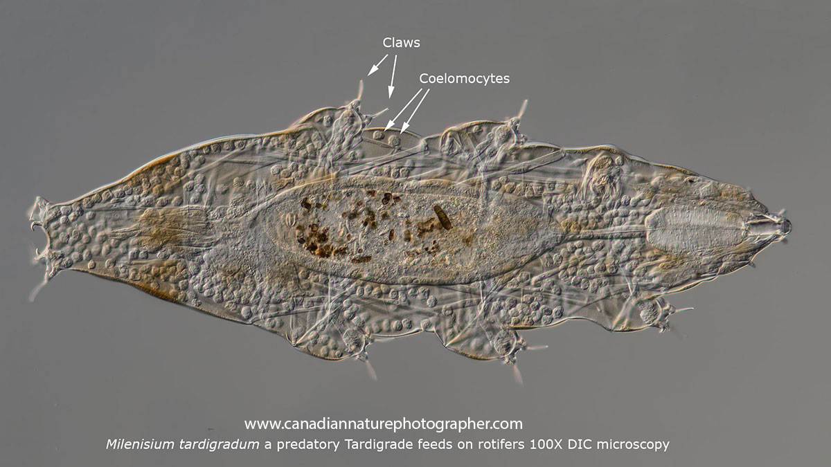
Milenisium tardigradum lives in lichen growing on a Mountain ash tree in my backyard. This tardigrade is a predator. DIC microscopy 100X.
Milenisium tardigradum predatory water bear DIC microscopy. Or you can view on YouTube https://youtu.be/Ih7BE5ZehjQ
Nematodes
Nematodes also called round worms are in the phylum Nematoda which includes more then 25,000 species. They are abundant in soil, water, eves troughs, moss cushions and pond water - almost every ecosystem on earth. On species, Caenorhabditis elegans is widely used in research to study cell lineage and neuronal development. Dr. Sydney Brenner proposed using this organism in research and it was the first multicellular organisms to have its whole genome sequenced, and in 2012 it had its neuronal "wiring diagram" completed. Dr. Brenner shared the Nobel prize in Physiology in 2002 for his work on this round worm. No organism that is studied carefully is insignificant and can contribute important information. Identifying the different species of nematodes is best left to experts as they are difficult to tell apart.
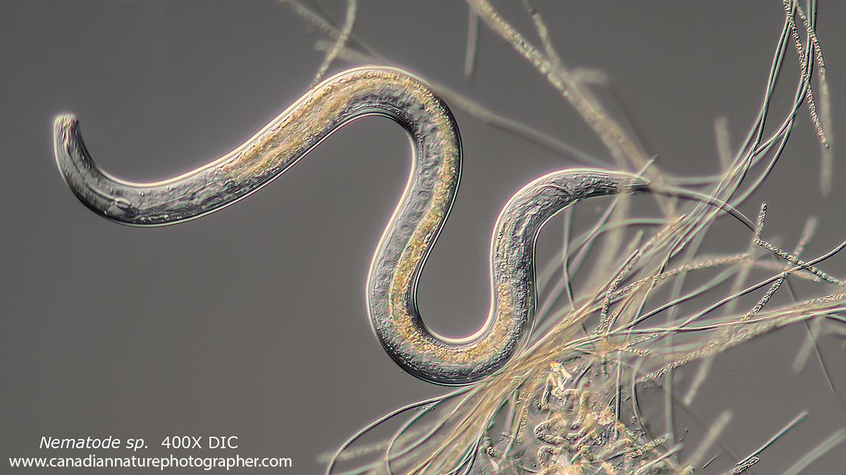
Nematode in pond water. DIC microscopy 400X.
Triclads or Flat worms
Triclads are flat worms also known generally as planarians. Triclads have a triple branched intestine (digestive gland). They are common in soil, ponds, and marine water. They are often found under rocks in water. They may have no eyes or many eyes on their head. Some species have auricles on the side of their head with mechanical and sensory receptors. Most are hermaphrodites. They have a remarkable ability to regenerate when cut. They have also been tested for their use in biological control of mosquitoes - they feed on the mosquito larvae.

Above Flatworm with 2 eyes and several eggs inside its body. Darkfield microscopy 40X
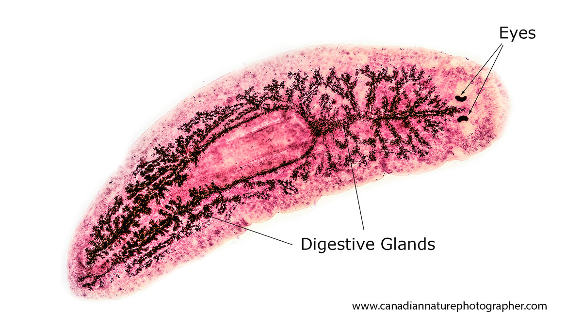
Above - Triclad that has been stained to show the triple branched digestive system after which they are named. 40X Brightfield microscopy.
Crystals and other Biological specimens that exhibit Birefringence
Birefringence is the optical property of a material having a refractive index that depends on the polarization and propagation direction of light. When polarized light goes through a specimen that exhibits birefringence it causes interference so that some colours (wavelengths) are removed from white light and the specimen appears coloured. Crystals viewed and photographed with a microscope and polarized light display the most intense and saturated colours I have ever seen in nature. Most biological specimens exhibit some birefringence under polarized light including muscles, hair and other structures where the molecules are arranged in an orderly pattern. Vitamin C crystals (ascorbic acid) are my favourite. To create these crystals I dissolve Vitamin C powder in water-isopropyl alcohol (1:1) then put the solution on a microscope slide and allow it to dry. The resultant crystals can resemble flowers and form a wide variety of geometric shapes. The British navy used Vitamin C stored as lemon and lime juice in barrels aboard their ships to give to sailors on long journeys especially journeys into the Arctic regions to ward off scurvy and hence British sailors became known as limeys. Indigenous people in North America avoided scurvy in winter by eating fresh meat and the Iroquois used a tea from the bark and leaves of an evergreen tree to ward off scurvy.
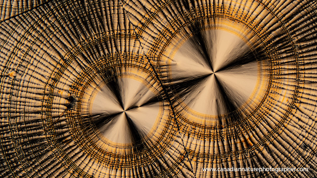
Vitamin C crystals by polarized light microscopy 100X

Vitamin C crystals polarized light microscopy about 50X
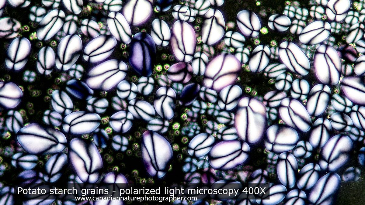
Potato starch grains in polarized light exhibit crystalline properties because how the starch molecules are arranged. The crystalline properties are lost after cooking. Different plants have characteristic starch grains. Polarized microscopy 400X. To see starch grains just squish a small piece of potato onto a microscope slide and view by polarized light microscopy. The easiest way to convert your light microscope to one with polarized light is shown in this YouTube video. You can also purchase linear polarizers and position one above the light source and one in the eyepiece for even better results.

A drop of water that has been frozen in my freezer on a microscope slide. The water when viewed by polarized light microscopy exhibits crystalline properties and displays the colours shown above. The frozen water will melt quickly on your microscope slide and you may only get a minute at most to observe these colours by polarized light microscopy.

Some of the most beautiful crystals of water are snowflakes. This one was photographed in my backyard. In order to take photos like this you need to catch the snowflakes on microscope slides that have been cooled outside for a couple of hours. I use macrophotography equipment and a light microscope to photograph snowflakes. I put the microscope in my garage which is at the same temperature as outside so the snowflakes won't melt for a minute or two - long enough to capture them with my camera. You can also preserve snowflakes permanently using super-glue.

Above shows a variety of new stereo-microscopes, light microscopes and magnifying lenses varying in price from $15 to $1000. You can save money by buying a used microscope though you need to do a bit more research or seek out the assistance of someone that is knowledgeable. To take pictures I would recommend a microscope with a trinocular head, though it is possible to get OK photos by simply holding your phone up to the eyepiece as shown in the photo below taken by students from Salisbury middle school.

The photo above was taken by students using a cell phone and a microscope provided by their school. If you are a student check if your school has microscopes and see if they will let you use one of them or get assistance from your science teacher. Alternatively consider purchasing your own microscope. It's unfortunate that libraries don't offer some microscopes to borrow, but microscopes are not hard find and are more affordable then ever. If you need help finding one I will be glad to point you to some sources.

Above - self portrait in my first microscope laboratory taken in my garage showing pictures of various micro-organisms on the wall.
Summary
Owning a microscope will help you discover an amazing universe that is all around you - the Micro-Universe. You can use a microscope in the comfort of your home all year long. Amateur astronomers need to find a dark site often far from city lights and it can get cold and lonely staying out until the early morning hours - I know because I used a telescope for several years as well.
I recommend microscopes to anyone interested in biology, nature, science or photography. They are fascinating to use whether you are young or retired and want to enhance your sense of wonder. If you need help getting started in microscopy feel free to contact me and if you live in the Calgary area I would be happy to offer you free advice, and make recommendations on where and what to buy to help get you started. RB
Author Biography & Contact Information
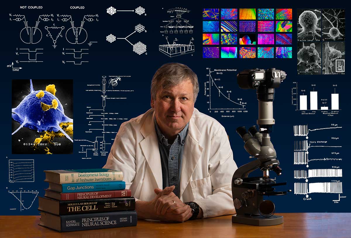
Bio: Robert Berdan is a professional nature photographer living in Calgary, AB specializing in nature, wildlife and science photography. Robert retired from Cell\Neurobiology research to take up photography full time years ago. Robert is an adjunct in the Dept. of Cell Biology at the University of Calgary. Robert offers photo guiding and private instruction in all aspects of nature photography and Adobe Photoshop training - including photomicrography and macrophotography. Portrait of Robert Berdan with pictures from some of his science publications in the background. Photo taken by Dr. Sharif Galal.
Email at: rberdan@scienceandart.org
Web sites:
www.canadiannaturephotographer.com
www.scienceandart.org
Phone: 9 am -7 pm MST (403) 247-2457.
Related Microscopy Articles by Robert Berdan on this web site
1. Photomicrography of Hydra - a model for studying regeneration and aging
2. Photographing Daphnia
3. Photographing Gastrotrichs
4. Photographing Rotifers
5. Photographing Ciliates
6. Photographing Stentors - A Large Unicellular Protozoan (ciliate) living in Freshwater
7. How to Collect and Photograph Water Bears (Tardigrades).
8. Tips on How to take Better pictures with a Microscope
9. Microscopic Pond Organisms from Silver Springs Calgary
10. Microscopic Life in Ponds and Rainwater - Pond Scum I
11. Photographing Microscopic Plant and Animal Life - Pond Scum II
12. Photomicrography and Video of Protozoa, Volvox and Rotifers
13. Home Microscopy Laboratory for Photomicrography
14. The Art & Science of Photomicrography with Polarized Light
15. Photographing Through a Microscope Photomicrography - Inner Space
16. Focus Stacking comparing Photoshop, Helicon Focus and Zerene
17. Rheinberg Filters for Photomicrography
18. Scanning Electron Microscopy - Photography
19. Photomicrographs of Diatoms from 1877 by John T. Redmayne
20. Microscopic Pond Life - Summer of 2019
Click on the buttons below and share this site with your friends