
Photographing Gastrotrichs
by Dr. Robert Berdan
July 28, 2018 (updated Dec 2019)

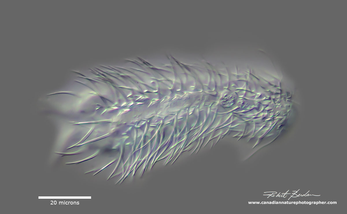
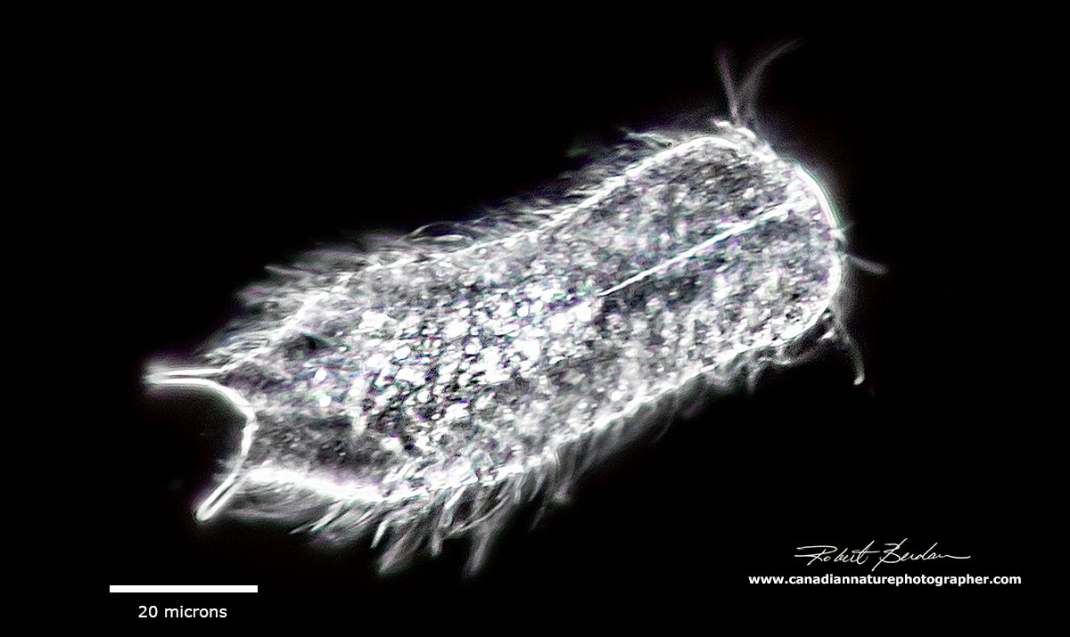
Ventral view of Gastrotrich - Chaetonotus sp. note the posterior caudal prongs and circular mouth near the apical end. Their shape is sometimes called "Tenpin" after the shape of bowling pins. Some species have many spines that are porcupine-like and most likely function to discourage predators.
Introduction to Gastrotrichs
To say that Gastrotrichs are a strange animal is an understatement. These tiny creatures vary in size from 60 to 3000 microns (micron= 1\1000 mm) though most found in Freshwater are 100-300 microns in size so you need a microscope to view and photograph them. There are 860 species worldwide, 355 species living in fresh water and more are being discovered. They are found living among the algae or along the bottom of ponds, lakes and streams and in the ocean. Supposedly they are the 5th most abundant microorganism in freshwater though I usually encounter them by accident while looking for other organisms.
The word gastrotrich comes from latin "stomach + hair". Some researchers refer to them as "hairy backs", but I think their spines resemble those of porcupine. They are pseudocoelomate animals - there is no body cavity. They are ciliated on their ventral surface, the cilia provide locomotion. In addition to numerous spines they have caudal fork at their posterior end which can consist of two to four prongs. Inside the prongs are adhesive glands - one to attach and one to dissolve the adhesive which I presume they use to attach to plants and other objects though I have never observed them attached to anything. On their head they have a circular mouth that opens into an elongated muscular pharynx. Whiskers on or around the mouth are cat-like in appearance and function as mechanoreceptors. The cuticle is thickened in regions and forms scales, hooks, and spines.
Freshwater species are parthenogenetic and produce eggs which are unfertilized. The eggs are about 50 microns in size and hatch into miniature adults. The eggs can sometimes be seen inside the animals. Gastrotrichs feed on detritus, organic materials, and bacteria. In turn they are preyed upon by turbellarians (flat worms) see below and carnivorous ciliates.

Flat worm that preys upon Gastrotrichs
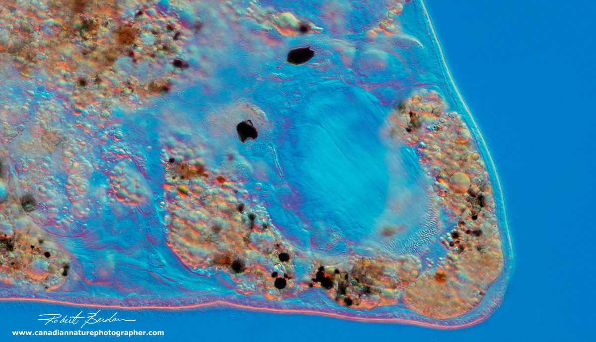
Enlargement of the head of a flat worm showing two black eye spots
Generally gastrotrichs live for only a few days. One species lived 40 days in a laboratory culture and produced 4-5 eggs. They produce two types of eggs, one which hatches quickly and a thicker shelled "resting" egg which is resistant to drying and freezing. Like rotifers, they exhibit eutely - they have a constant number of cells. They have a brain (cerebral ganglion) which leads to a pair of nerve cords along either side of the body. Their primary sense organs are bristles and ciliated tufts which function as mechanoreceptors. Some gastrotrichs also possess light sensitive cells in the brain that work as primitive ocelli (eye spots) though I have never seen any.
The animals move in a gliding action, not as fast as some ciliates, but unless pinned down by an overlying coverslip they are in constant motion making photography of these animals challenging. (Freshwater species can be narcotized using 1% Magnesium Chloride Todaro et. al 2019). The size of the animals is an important characteristic and the number of spines is constant, though they will sometimes break a spine and it will not regenerate. While the number of spines might be taxonomically important I challenge anyone to count them while this animal is moving - I presume counting spines can only be achieved in specimens that are fixed or narcotized. One method to collect these animals is to get handfuls of algae and water plants and squeeze them into a container - though you will also collect lots of other organisms like rotifers, daphnia etc doing this.

Diagram of Gastrotrich
Taxonomy
Kingdom: Animalia
SuperPhyllum: Rouphozoa The roufozoos (Rouphozoa) are a clade ( organisms believed to have evolved from a common ancestor) of animals that contain two edges, the one of flat worms (Platyhelminthes) and the one of thorny worms (Gastrotricha).
Phylum: Gastrotricha
Order: Chaetonotida - contains mostly freshwater, but some marine species, posses scales in addition to spines. 5 Families, 23 genera in freshwater.
Order: Macrodasyida - mostly marine, but a few freshwater species. Marine species are mostly found between sand grains.
For a Key to Freshwater Gastrotrich species see the link to Edmondson Fresh water biology - below - it is an online book. Also see the links below for more information on Gastrotrichs e.g. Todaro et al 2019.

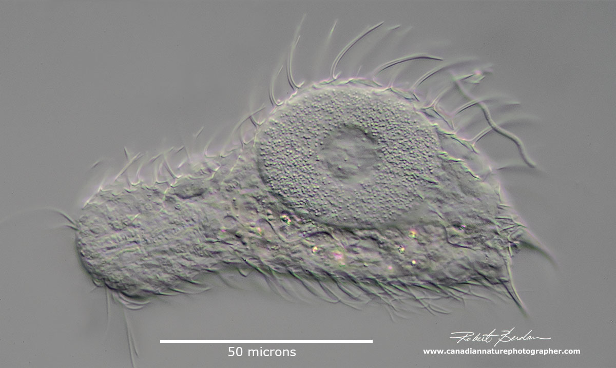
Chaetonotus sp Ventral view of the animal. Spines are the hallmark for species of Chaetonotus.

Chaetonotidus sp - DIC microscopy with Darkfield effect
Chaetonotus sp. side view of the animal DIC microscopy
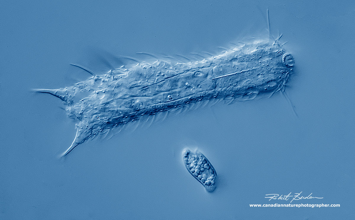
Chaetonotus sp. and a smaller flagellate - Euglenoid sp.

Chaetonotus sp. and two smaller flagellates - Euglenoid sp.
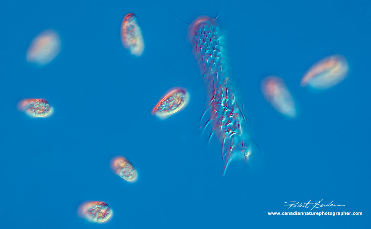
Dorsal view of Chaetonotus sp. showing its many spines and some smaller flagellates - Euglenoid sp.
Chaetonotida sp - I focused on the dorsal surface to show the porcupine-like spines.

Chaetonotida sp - I focused on the dorsal surface to show the porcupine-like spines.

Chaetonotida sp - I focused on the dorsal surface to show some of the scales

Lepidodermella squamata and a Diatom (Pinnularia nobilis) Phase contrast microscopy 400X

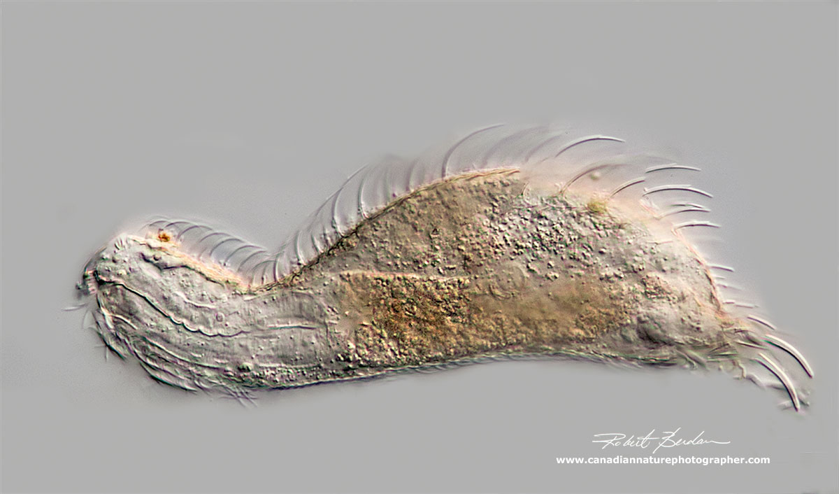
Lepidodermella squamata (Order: Chaetonotida, Family: Chaetonotidae) there are two adhesive suckers on the posterior end. The surface of the trunk is covered in scales. There are tufts of bristles on the head and two rows of cilia on the ventral surface of the body. DIC microscopy.
Diagram showing some different Freshwater Gastrotrichs
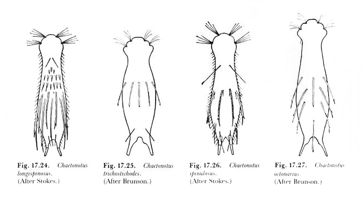
From Edmondson (1959) page 416
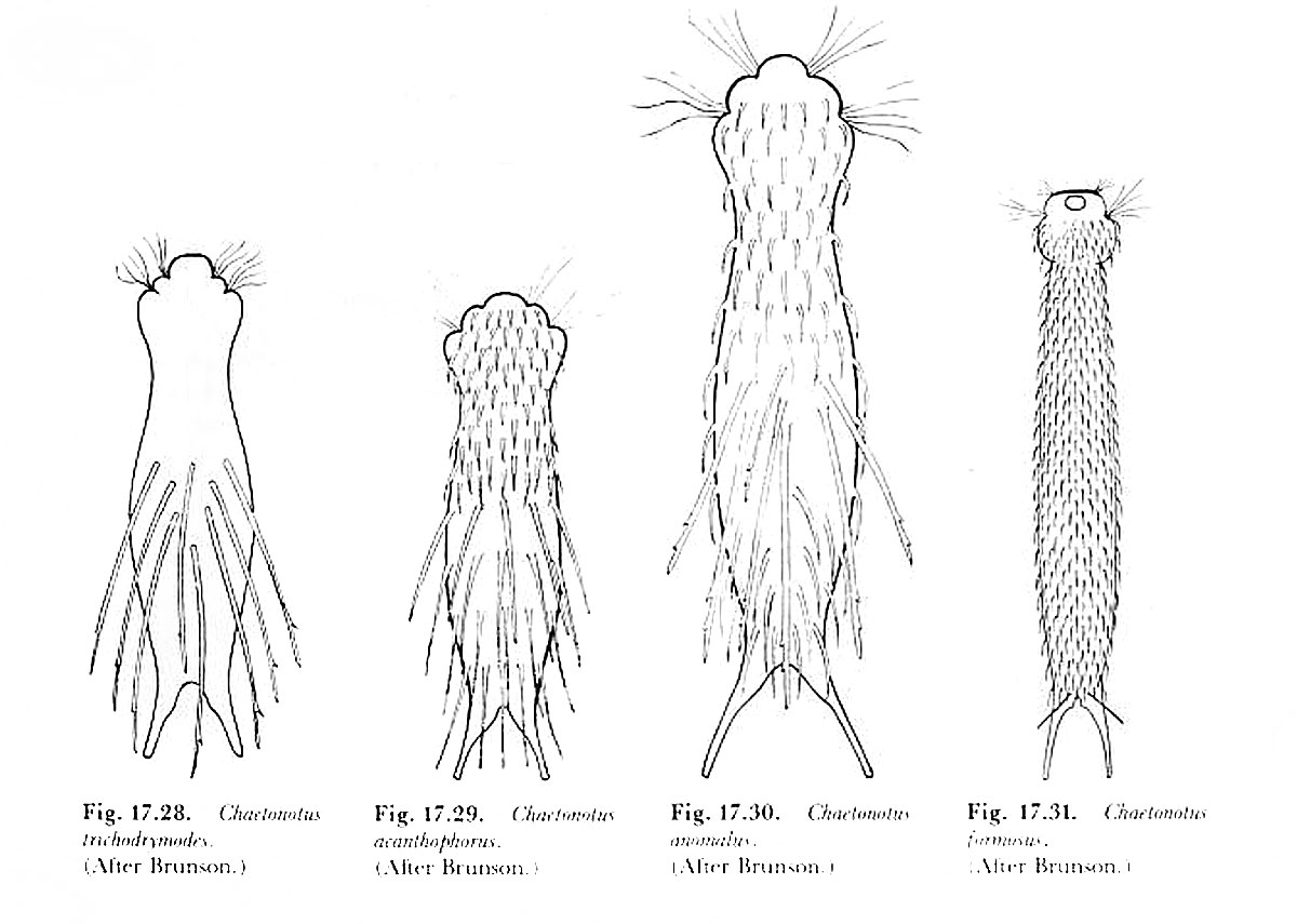
From Edmondson (1959) page 417
Examples of More species of Gastrotrichs from a recent research paper
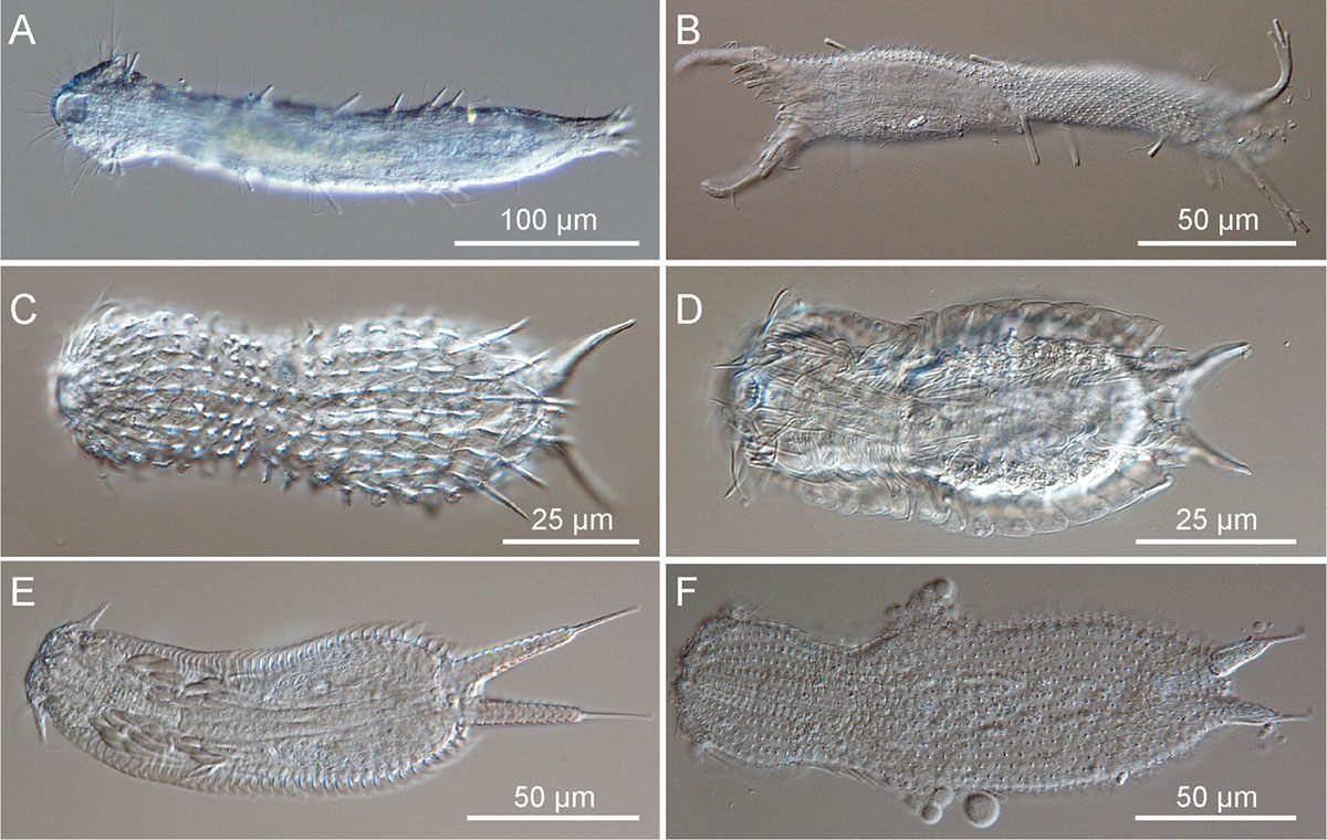
A & B = Macrodasyida C, D, E & F = Chaetonotida M. Antonio Todaro, Matteo Dal Zotto, Sarah J. Bownes, Renzo Perissinotto - Todaro, M. A., Dal Zotto, M., Bownes, S. J., Perissinotto, R. (2011). First records of Gastrotricha from South Africa, with description of a new species of Halichaetonotus (Chaetonotida, Chaetonotidae). From ZooKeys 142: 1–13 - Wikipedia.

DIC microscopy (A, C) and SEM (scanning electron microscopy) (B, D, E) photomicrographs showing the general body shape and aspects of the cuticular covering of representatives of the genera Pseudostomella and Ptychostomella. Scale bars A, C = 50 µm, B, D, E = 20 µm. From Wikipedia - authors: M. Antonio Todaro, Tobias Kånneby, Matteo Dal Zotto, Ulf Jondelius. PLoS ONE 6 (3): e17892. doi:10.1371/journal.pone.0017892

Lepidodermella squamata by DIC microscopy - these animals are very flexible and can turn their bodies almost 180 degrees to form a U shape.
Lepidodermella squamata - viewed by Darkfield microscopy. Note the caudal prongs.

Lepidodermella squamata - DIC microscopy.
Gastrotrich with egg inside
Summary
Freshwater Gastrotrichs are microscopic in size (100-300 microns) and found mainly around water plants or the benthos (bottom of a body of water). They are often tenpin shaped like a bowling pin, have ventral ciliation usually in two bands. The have a terminal or subterminal mouth connected to a muscular sucking pharynx and feed mostly on detritus, bacteria, small protozoa, and algae. They have a posterior fork-like structure called a furca that bears adhesive glands. Although they are not abundant in my experience they are common enough that anyone looking at pond water with a microscope will encounter them sooner or later. Sooner if you squeeze some of the algae to extract them.
Photomicrography
All photographs my photographs are of live Gastrotichs collected from fresh water water near my home. For details on how I took these pictures please see my article - Tips on How to Take Better Pictures with a Microscope - Photomicrography. In brief, I used a Nikon D500 and Nikon D800 cameras, Zeiss Axioscope with DIC, Bright field, and Darkfield lighting, an Olympus E microscope equipped with Phase contrast and controlled my cameras with free software Digicam control. I slowed them down for photography by mild compression under the coverslip.
References:
(2019) M.A. Todaro et. al. An Introduction to the Study of Gastrotricha, with a Taxonomic Key to Families and Genera of the Group. Diversity, 11, 117 - Open Access PDF
(2019) Gastrotricha Web Portal - http://www.gastrotricha.unimore.it/
Dr. M.A. Todaro web site in Italy - http://www.gastrotricha.unimore.it/todaro.htm
(2015) Kanneby. T, Hochberg, R. In Thorp and Covich's Freshwater Invertebrates: Ecology and General Biology. Thorp, J., Rogers, D.C., Eds Elsevier Academic Press: Amsterdam, The Netherlands Vol i. pp. 211-223.
(2011) M. Antonio Todaro , Tobias Kånneby, Matteo Dal Zotto, Ulf Jondelius. Phylogeny of Thaumastodermatidae (Gastrotricha: Macrodasyida) Inferred from Nuclear and Mitochondrial Sequence Data. PLOS one - download PDF
(2011) T. Kånneby Gastrotricha of Sweden Biodiversity and Phylogeny. Dept. Zoology, Stochholm University Doctoral dissertation - Download PDF
(2010) T. Kånneby and R. Hochberg. Phylum Gastrotricha. Chapter 12 in Thorp and Covich's Freshwater Invertebrates 4th ed. pp 211-223. Academic Press, NY.
(2010) D. Strayer, W.D. Hummon, R. Hochberg. Gastrotricha Chapter 7 Ecology and Classification of North American Freshwater Invertebrates. Pg 163-172 - Download PDF
(1959) W.T. Edmondson (ed) Fresh Water Biology 2nd edition, Gastrotricha by R. B. Brunson. John Wiley. NY pg 406-419 - read online version free.
Note to teachers and students you have permission to use my pictures for teaching or school projects. I appreciate credit and\or link back to my web site if possible. For commercial use of my photos please contact me.
Authors Biography & Contact Information

Robert Berdan is a professional nature photographer living in Calgary, AB specializing in nature, wildlife and science photography. Robert retired from Cell\Neurobiology research to take up photography full time years ago. Robert offers photo guiding and private instruction in all aspects of nature photography and Adobe Photoshop training - including photomicrography, macrophotography.
Related Microscopy Articles by Robert Berdan on this web site
1. Photographing Rotifers
2. Photographing Ciliates
3. Photographing Stentors - A Large Unicellular Protozoan (ciliate) living in Freshwater
4. How to Collect and Photograph Water Bears (Tardigrades).
5. Tips on How to take Better pictures with a Microscope
6. Microscopic Pond Organisms from Silver Springs Calgary
7. Microscopic Life in Ponds and Rainwater - Pond Scum I
8. Photographing Microscopic Plant and Animal Life - Pond Scum II
9. Photomicrography and Video of Protozoa, Volvox and Rotifers
10. Home Microscopy Laboratory for Photomicrography
11. The Art & Science of Photomicrography with Polarized Light
12. Photographing Through a Microscope Photomicrography - Inner Space
13. Focus Stacking comparing Photoshop, Helicon Focus and Zerene
14. Rheinberg Filters for Photomicrography
15. Scanning Electron Microscopy - Photography
16. Photomicrographs of Diatoms from 1877 by John T. Redmayne
Email at: rberdan@scienceandart.org
Web site: www.canadiannaturephotographer.com
Phone: MST 9am -7 pm (403) 247-2457.
Click on the buttons below and share this site with your friends